Targeted nutritional intervention für down-syndrom
Targeted Nutritional Intervention für
Down-Syndrom
Vortrag anlässlich der insieme 21 Tagung mit Schwerpunktthema
Ernährung und Nahrungsergänzung in Zug am 15.11.08
(Letzte Aktualisierung: November 2010)
Autor: Richard Müller, Zürich
• Was ist TNI?
• Ginkgo Biloba, Fischöl, Probiotika, Vitamin D und Curcumin
• Warum TNI, wenn mein Kind doch gesund ist?
• Gibt es wissenschaftliche Untersuchungen, die TNI rechtfertigen?
• Wer stellt TNI Produkte her? Gibt es eine Qualitätskontrolle?
• Ist mit Nebenwirkungen zu rechnen?
• Wo sind TNI Produkte erhältlich und wieviel kosten sie?
Informationsquellen
• Bibliographie
• Anhang (TNI bei Kindern mit Down-Syndrom)
Was ist TNI?
Wir sind es gewohnt, beim Down-Syndrom im Allgemeinen über Herzfehler,
Fehlfunktionen des Verdauungstraktes oder der Schilddrüse zu sprechen, also
Störungen auf der Organebene. Diese lassen sich in den meisten Fällen heute
gut behandeln. Über Störungen der Abläufe innerhalb der einzelnen Zellen
hören wir aber praktisch nie etwas. Diese lassen sich jedoch ebenfalls gut
behandeln.
Veränderte Stoffwechselbedingungen bei Menschen mit Down-Syndrom sind
in der wissenschaftlichen Fachwelt seit langem bekannt (1) - ganz im
Gegensatz zur Ärzteschaft, die in der Regel wenig oder nichts darüber weiss.
Es besteht offenbar zwischen dem Wissen in der Fachwelt und dem
Erfahrungswissen der behandelnden Ärzte – bis auf wenige Ausnahmen - eine
grosse Diskrepanz.
Das Ziel dieser vorliegenden Arbeit ist es, interessierten Eltern und
Fachpersonen im deutschsprachigen Raum einen Einblick in den gegen-
wärtigen Forschungsstand bezüglich dieses Themas zu ermöglichen.
Es ist leider so, dass die veränderten Stoffwechselabläufe beträchtliche
Auswirkungen auf den Körper und Geist von Menschen mit Down-Syndrom
haben. Einerseits kommen gehäuft Mangelzustände bei bestimmten
Nährstoffen vor und andererseits sind die Anzeichen für den sogenannten
oxidativen Stress erhöht (2). Da der Begriff „oxidativer Stress" einigen
Lesern nicht geläufig sein mag, wird er später im Detail erläutert. Jedenfalls
beeinflusst dieser oxidative Stress das Immunsystem und das Gehirn von
Kindern und Erwachsenen mit Down-Syndrom in negativer Weise (3, 4, 5).
Aufgrund des Nährstoffmangels und des erhöhten oxidativen Stresses ist eine
gezielte Nährstoffzufuhr angezeigt, die allein durch eine ausgewogene und
gesunde Ernährung nicht genügend gewährleistet werden kann.
TNI bedeutet „Targeted Nutritional Intervention" oder in Deutsch „gezielte Ernährungsintervention". Konkret ist das der Versuch, die gesunde Balance des gestörten Stoffwechsels wiederherzustellen.
Dabei muss klar sein, dass TNI das Down-Syndrom keineswegs heilt, es ersetzt auch keine anderen Massnahmen der Förderung von Menschen mit Trisomie 21.
Das TNI-Behandlungsprotokoll besteht aus sechs separaten Bestandteilen. Einerseits aus einem kommerziell erhältlichen Produkt, das Vitamine, Mineral-
1 So wird etwa der oxidative Stress bei einem 10 kg schweren Kind mit DS erst mit einer
Menge von antioxidativem CoenzymQ10 neutralisiert, die einem halben Kilogramm Schweineherz pro Tag entspricht (6a, 6b)!
stoffe, bestimmte Aminosäuren, einige Pflanzenwirkstoffe und Verdauungs-enzyme enthält, und andererseits kommen zusätzlich Omega-3-Fettsäuren in Form von Fischöl hinzu, ausserdem Ginkgo Biloba, ein Probiotikum, Vitamin D und Curcumin.
Wie allgemein bekannt, wird das Down-Syndrom durch das überzählige dritte Chromosom 21 in jeder Körperzelle verursacht. Es handelt sich also um einen Überschuss - es ist gleichsam etwas zuviel vorhanden. Man könnte meinen, dass das nicht so schlimm ist. Es fehlt ja nichts, im Gegenteil. Gewisser-massen ist das auch richtig: wäre nämlich ein Chromosom zuwenig vorhan-den, könnte ein Mensch gar nicht leben. Trotzdem verursacht aber ein Zuviel an Genmaterial einen ziemlichen Schaden. Warum? Nun, auf den Chromo-somen ist das menschliche Erbgut in Form von Genen gespeichert. Das heisst also, dass ein Chromosom zuviel auch ein Zuviel an Genen bedeutet. Unsere Gene sind in jeder Körperzelle aktiv, indem sie Eiweisse produzieren wie zum Beispiel Enzyme. Dies trifft ebenfalls auf das 21. Chromosom zu. Heute weiss man, dass auf Chromosom 21 Gene liegen, die das Enzym Superoxid-Dismutase und Cysthathionin-Beta-Synthase produzieren (5, 7). Bei Menschen mit Down-Syndrom sind diese beiden Enzyme also in 50% erhöhter Anzahl vorhanden, und dies stellt ein grosses Problem dar. Warum ist das so? Nun, beide Enzyme sind aktiv am Stoffwechsel jeder Körperzelle beteiligt und bei Menschen mit Down-Syndrom tun sie dies daher – weil in erhöhter Anzahl vorhanden - auf überaktive Weise (8, 9). Was das konkret bedeutet, wird im Folgenden anhand von einigen schematischen Darstellungen aufgezeigt. Gleich anschliessend ist ein Ausschnitt der Stoffwechselkreisläufe veranschaulicht, an denen das Enzym Cysthathionin-Beta-Synthase beteiligt ist (10). Prominent in der Mitte erscheinen zwei Kreisläufe, die auf den ersten Blick völlig normal verlaufen. Auch die beiden erwähnten Enzyme sind einge-tragen; eines davon ist gleich unterhalb des grossen Kreislaufs in der Mitte ersichtlich. Es ist eingekreist und trägt das Kürzel „CBS", was für Cysthathionin-Beta-Synthase steht. Der kleine aufwärtsgerichtete Pfeil neben CBS bedeutet, dass das Enzym überaktiv ist, da es ja dreimal anstatt nur wie normalerweise im Doppel vorhanden ist. Dies wird in der schematischen Darstellung verdeutlicht: die zwei grünen Pfeile zeigen eine normale Aktivität; der zusätzliche rote Pfeil veranschaulicht, dass beim Down-Syndrom ein zusätzliches CBS Enzym aktiv ist.
Das hat zur Folge, dass 50% zuviel Homocystin aus dem Kreislauf „nach unten" herausgezogen wird. Homocystin ist eine wichtige Aminosäure - in der Darstellung gleich oberhalb des CBS-Enzyms vermerkt. Der abwärts gerichtete Pfeil neben dem Wort Homocystin bedeutet übrigens genau das, nämlich dass dem Stoffwechselkreislauf oben zu wenig Homocystin zur Verfügung steht. Dies hat bedeutet schliesslich, dass die Protein-Synthese in
der Zelle gestört ist (rechts aussen: Protein SynthesisÈ). Protein-Synthese bedeutet die Produktion von Eiweissen, welche die Voraussetzung dafür ist, dass sich die Körperzellen teilen können, was wiederum die Grundlage für das Wachstum des Körpers ist. Auch der nachbarliche Stoffwechselablauf wird durch den Homocystin-Mangel in Mitleidenschaft gezogen. Tetrahydrofolat, abgekürzt THF, eine Form des B-Vitamins Folsäure, ist dadurch ebenfalls reduziert. Aufgrund dieses Mangels ist die DNA-Synthese, als auch die Produktion der Erbsubstanz und von Neurotransmittern, den Gehirnbotenstoffen, reduziert (11, 12, 13). Auch das hat einen nachteiligen Effekt auf die Körperzellen, da sie sich nicht mehr gut reparieren können; sie altern und sterben daher schneller. Bei Menschen mit Down-Syndrom lässt sich dies übrigens gut feststellen: in der Regel ist das Körperwachstum reduziert und der Alterungsprozess beschleunigt. Überdies
korreliert der Folsäure- und Homocystinstatus von Menschen mit Down-Syndrom unabhängig von der Altersgruppe stark mit dem Intelligenz-quotienten (14). Bei der Betrachtung obiger Darstellung fällt auf, dass häufig Vitamine bei den Stoffwechselkreisläufen beteiligt sind. In diesem Fall handelt es sich vor allem um die Gruppe der B-Vitamine, speziell um Vitamin B12 (Cobalamin) und B9 (Folsäure). Wie lässt sich diese Stoffwechselstörung nun durch TNI beeinflussen? Durch die erhöhte Zufuhr von Vitamin B12 und von Folinsäure, einer speziell gut bioverfügbaren Form von Folsäure (15), kann Kreislauf links im Bild, der Folsäurestoffwechsel gestärkt werden (7). Die Folge davon ist, dass mehr Homocystin von den CBS Enzymen gleichsam weggezogen und dem Stoffwechselkreislauf rechts zugeführt wird. *** Cysthathionin-Beta-Synthase ist nicht das einzige Enzym, das beim Down-Syndrom verdreifacht ist. Es gibt noch eines, das auf dem 21. Chromosom genetisch gespeichert ist, und der Schaden, den es anrichtet ist ungleich dramatischer als bei CBS; es handelt sich um Superoxid-Dismutase (links unten in obiger Darstellung: SOD) (16). Dieses Enzym ist mit verantwortlich dafür, dass Menschen mit Down-Syndrom schneller altern (17, 18, 19), dass die kognitiven Fähigkeiten mit der Zeit nachlassen und sie vermehrt an Alzheimer erkranken (5, 20, 21). Der Mechanismus, über welchen dieses Enzym Schaden verursacht, hängt mit der Produktion sogenannter freier Radikale zusammen. Freie Radikale entstehen ständig beim ganz normalen Stoffwechsel in der Zelle. Was sind freie Radikale eigentlich? Es sind Moleküle, die ein Elektron verloren haben, z.B. durch UV-Licht, Umweltgifte oder natürliche Stoffwechselvorgänge innerhalb der Zellen. Das fehlende Elektron macht sie zu aggressiven Zellgiften, die grossen Schaden in der Zelle anrichten können, wenn sie nicht sofort neutralisiert werden. Sie versuchen nämlich bildlich gesprochen das fehlende Elektron aus anderen Strukturen innerhalb der Zellen herauszu-reissen, um wieder vollständig zu werden. Freie Radikale entstehen ständig – jede einzelne unserer Körperzellen wird durchschnittlich circa zehntausend Mal pro Tag von Freien Radikalen angegriffen!
Unsere Zellen sind umgeben von einer Membran, die aus zwei dünnen, flexiblen Lagen aus Fettmolekülen besteht, welche in der nachfolgenden Abbildung Phospholipide genannt werden. Es handelt sich nicht um irgend-welche Fette, sondern um Omega-3-Fettsäuren, die mit der Nahrung, speziell
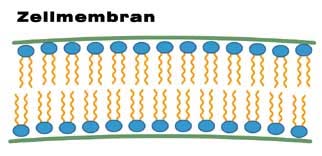
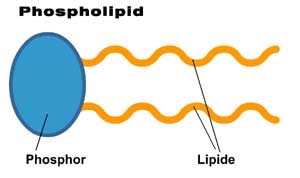
mit fetthaltigem Fisch, aufgenommen werden. Diese fetthaltige Zell-membran ist sehr delikat und leicht verletzbar, und daher stellen die aggressiven freien Radikale ein grosse Bedrohung für sie dar. Die zwei Fettlagen, welche die Zelle gegen innen und aussen abdichten schützen ihr Inneres, insbesondere die Erbanlagen im Zellkern. Rechts im Bild ist ein Einzelteil, ein Phospholipid im Grossformat zu sehen. Die gelben Wellenlinien stellen die gegenüber Oxidationsprozessen empfindlichen Omega-3-Fettsäuren dar.
Innerhalb der Zelle laufen während der Energieproduktion natürliche Stoffwechselvorgänge ab, welche eine Vielzahl von Freien Radikalen produzieren. Die Freien Radikale gleichen dann sozusagen den Auspuffgasen dieser Energieproduktion. Damit diese Abfallprodukte entsorgt werden können, stehen den Zellen sogenannte Radikalfänger zur Verfügung, welche die Aufgabe haben, die Freien Radikale unschädlich zu machen. Es handelt sich sowohl um körpereigene Radikalfänger, bestimmte Enzyme, als auch solche, die von aussen mit der Nahrung zugeführt werden. Diese heissen Antioxidantien, und dabei handelt es sich vor allem um die Vitamine A, C und E als auch um bestimmte Spurenelemente, allen voran Selen.
In einer gesunden Zelle herrscht eine fein abgestimmte Balance zwischen der Zahl von Freien Radikalen und den Radikalfängern. In den Zellen von Menschen mit Down-Syndrom ist diese Balance jedoch bereits vorgeburtlich (22) und über das gesamte Lebensalter hin empfindlich gestört (23, 24, 25). Und hier kommt das vorhin erwähnte Enzym Superoxid-Dismutase zum Zug. Dieses Enzym ist eigentlich ein geniales kleines Ding, da es eines der wichtigsten körpereigenen Radikalfänger ist. Was macht es? Es „dismutiert" oder verwandelt das giftige Superoxid-Radikal zu Wasserstoffperoxid, das weniger giftig ist. Mit der folgenden Darstellung lassen sich diese Vorgänge
etwas leichter vorstellen. Mit „Respiratory Chain" (links) wird die Energieproduktion der Zelle ange-deutet. Daraus entsteht sozusagen als Auspuffgas das Freie Sauerstoff-Radikal O2-Minus, bzw. Superoxid.
ieses wird durch das Enzym Superoxid-Dis
mutase (SOD) zu H2O2 oder
Wasserstoffperoxid umgewandelt. Wasserstoffperoxid ist allgemein als Bleichmittel bekannt, das man zum Blondfärben der Haare verwendet. Es handelt sich um eine ziemlich aggressive Substanz, wie man sich denken kann. Dieses wird anschliessend durch ein weiteres Enzym namens Catalase in harmloses reines Wasser verwandelt.
Man mag sich nun fragen, wo hier das Problem ist. Es funktioniert doch alles
bestens, viel besser jedenfalls, als wir Menschen irgendwelche Umweltgifte
unschädlich machen können. Nun, das Problem von Zellen mit Trisomie 21 ist
folgendes: Das Enzym Catalase ist auf dem Chromosom 11 gespeichert, und
zwar wie es von der Natur vorgesehen wurde in zweifacher Ausführung. Das
Enzym Superoxid-Dismutase ist jedoch auf Chromosom 21 gespeichert, also
ist es verdreifacht, eben nicht, wie es von der Natur vorgesehen wäre. Wir
haben also drei Enzyme Superoxid-Dismutase, die „eifrig vor sich
hinwerkeln", indem sie Wasserstoffperoxid produzieren und nur zwei Exem-
plare von Catalase, die hinterher sauber machen. Diese Rechnung kann so
nicht aufgehen (26).
In der nachfolgenden schematischen Darstellung wird der Unterschied zwischen einer gesunden Zelle und einer Trisomie-Zelle gezeigt. Bei beiden wird Superoxid zu Wasserstoffperoxid umgewandelt, aber bei Trisomie 21 wird zuviel Wasserstoffperoxid produziert und es entsteht ein Überschuss,
der von den nachfolgenden Enzymen nicht vollständig abgebaut werden kann. Es kommt leider noch schlimmer, wie unten aufgezeigt wird. Aus dem überschüssigen Wasserstoffperoxid wird das Freie Radikal OH-Minus, Hydroxyl gebildet, speziell bei Vorhandensein von Eisen („Fenton Reaktion") (27). Bei Hydroxyl handelt es sich um eines der schlimmsten Zellgifte, die es gibt.
Die aggresive oxidative Aktivität der Hydroxyl-Radikale führt zur Schädigung der Erbanlagen innerhalb des Zellkerns (mtDNA/mitochondrial DNA) und an der Peripherie der Zelle zur Lipidperoxidation, das heisst zur Zerstörung der Fettsäuren in den Zellwänden. Schliesslich wird dadurch ein Teufelskreis in Gang gesetzt, durch den immer grössere Schäden entstehen, wie in der
nächsten Illustration gezeigt wird.
Links aussen greifen Freie Radikale - wie zum Beispiel das Hydroxyl-Radikal - eine Zelle an, was zu Schäden an den delikaten Omega3-Fettsäuren in den Zellwänden führt. Aus den Bruchstücken entstehen entzündungsfördernde Substanzen, die ihrerseits wieder Freie Radikale erzeugen und so weiter.
Es gibt bereits zahlreiche wissenschaftliche Studien, die bei Menschen mit Down-Syndrom diese erhöhte Lipidperoxidation nachweisen (19, 23). Dies lässt sich relativ einfach mit einem Bluttest bestätigen (Plasma- und Erythrozyten Malondialdehyd-Werte). Diese Schäden entstehen bereits, während sich das ungeborene Kind im Mutterleib befindet (22).
Was sind denn nun die konkreten Folgen dieser Schäden? Hat die Zellwand, dieser erste Schutzwall um die Zelle herum erst einmal Schaden genommen, können Freie Radikale ungehindert in die Zelle eindringen und bis zum Zellkern gelangen. Genau genommen bis in den Zellkern hinein, wo sie die Erbanlagen schädigen. Ist die Erbsubstanz erst einmal geschädigt, wird sich diese Zelle in alle Zukunft nur noch beschädigt reproduzieren können. Unsere Zellen altern, werden teilweise repariert, müssen aber auch immer wieder ersetzt, d.h. durch Teilung erneuert werden. Bei diesem Vorgang wird eine genaue Kopie erstellt. Ist die Vorlage für die Kopie aber nicht mehr optimal, werden auch alle weiteren Kopien fehlerhaft sein. Dazu kommt, dass sich Zellen nicht unbegrenzt teilen können. Sind die Zellen also fehlerhaft erneuert worden oder können sie sich nicht mehr teilen bzw. erneuern, kommt es zu dauerhaften ‚Fehlern im System'. Die fehlerhaften Zellen lassen
den Organismus schneller altern. Fallen nur einzelne Zellen aus, fällt dies noch relativ wenig ins Gewicht. Bei zunehmender Anzahl werden jedoch die Funktionen beeinträchtigt – zuerst von Zellverbänden, dann von Organen, schließlich vom gesamten Körper. Das wohl empfindlichste Organ im menschlichen Körper ist das Gehirn. Und es ist das Gehirn von Menschen mit Down-Syndrom, das am meisten unter oxidativen Stress leidet (28). In Forscherkreisen wird daher das Down-Syndrom mitunter auch als Krankheit bezeichnet, weil es sich dabei unter anderem als eine fortschreitende Degeneration des Gehirns handelt (29, 30, 31, 32a, 32b). Die gute Nachricht bei alldem ist, dass etwas dagegen unternommen werden kann. Wissenschaftliche Studien haben gezeigt, dass dieser übermässige oxidative Stress bei Menschen mit Down-Syndrom neutralisiert werden kann. Die Verwendung therapeutischer Dosen von Antioxidantien (20, 16), wie Coenzym Q10 (6a, 6b), Vitamin A (33), C (34), E (35, 36) und Selen (37) ist
angezeigt. Diese gehören daher immer zu den Hauptbestandteilen der
verschiedenen auf dem Markt erhältlichen TNI-Produkte. Allein durch die
Ernährung kann der Bedarf an Antioxidantien für Menschen mit Down-
Syndrom nicht gestillt werden.
Ginkgo Biloba, Fischöl, Probiotika, Vitamin D und Curcumin
Aus der Down-Syndrom Hirnforschung ist seit Neuerem bekannt, dass die
Langzeitpotenzierung, eine wichtige Voraussetzung für das Lernen und das
Gedächtnis, bei Menschen mit DS stark eingeschränkt ist und dass diese
Veränderung durch eine erhöhte Aktivität der inhibitorischen (hemmenden)
Signalübertragung zwischen den Hirnzellen verursacht wird.
Langzeitpotenzierung heisst, dass Verbindungen zwischen den Hirnzellen
verstärkt (potenziert) werden, wenn derselbe Reiz immer wieder über die
gleiche Verbindung geleitet wird. Auf diese Weise wird das Abspeichern von
Gedächtnisinhalten, bzw. Lernen überhaupt erst ermöglicht.
In welche Richtung zielt die DS-Forschung aufgrund dieser Erkenntnisse?
Es ging im Anschluss an diese Entdeckung vornehmlich darum
herauszufinden, warum die inhibitorischen Signale bei DS überaktiv sind und
wie man diese Überaktivität medikamentös reduzieren könnte. Die
inhibitorische Signalübertragung ist grundsätzlich ein Segen, denn sie
bewirkt, dass der grösste Prozentsatz an Sinnesreizen, die auf das Gehirn von
aussen einstürmen als unbedeutend erkannt und herausgefiltert oder eben
gehemmt wird. Bildlich gesprochen werden diese hemmenden Rotlichter nur
für ganz wenige als wichtig eingestufte Reize auf Grün geschaltet, damit sie
passieren können. Nur diese auserwählten Reize werden anschliessend von
Hirnzelle zu Hirnzelle weitergeleitet, was zur Folge hat, dass sie als
Gedächtnisinhalte abgespeichert werden. Bei Menschen mit DS sind diese
Rotlichter überaktiv, was bewirkt, dass zu wenige Informationen weitergeleitet werden. In einer Studie mit DS-Mäusen wurde bewiesen, dass diese Rotlichter medikamentös auf Grün geschaltet werden können (38). Die dabei verwendete Substanz, Picrotoxin, schaltete aber zu viele Rotlichter auf Grün, was bewirkte, dass das Gehirn von Reizen überflutet wurde. Es versteht sich von selbst, dass Picrotoxin als Medikament für die Anwendung beim Menschen nicht in Frage kommt. Die Forscher suchen nun fieberhaft nach anderen Substanzen, welche die Eigenschaften von Picrotoxin in abgeschwächter Form aufweisen. Ein vielversprechender Kandidat aus einer „unerwarteten Ecke" ist ein Pflanzenheilmittel, das Extrakt der Blätter des Ginkgo-Baumes, welches in Asien bereits seit Jahrtausenden zur Verbesserung der Hirnleistung eingesetzt wird. Tatsächlich wurde in einer weiteren Studie (39) bereits nachgewiesen, dass die molekulare Struktur des relevanten Ginkgo Biloba Wirkstoffes (Bilobalid) fast haargenau derjenigen von Picrotoxin gleicht, nur schaltet offenbar Bilobalid nur geradeso viele Rotlichter auf Grün wie nötig. Klinische Studien an Menschen mit DS, um die Wirksamkeit von Ginkgo Biloba als Verstärker der Langzeitpotenzierung zu testen sind bereits in Planung (Stand: 2008). Da bei den Down-Syndrom Mäusen aber derart signifikant gute Resultate erzielt wurden und klinische Studien noch lange auf sich warten lassen, entscheiden sich immer mehr Eltern, ihren Kindern mit DS Ginkgo Biloba zu verabreichen, da es als Naturheilmittel rezeptfrei erhältlich ist und eine ausgezeichnete Verträglichkeit aufweist. Aus diesen Gründen wurde dem TNI-Protokoll im Jahr 2007 Ginkgo Biloba hinzugefügt (Empfehlung: erst ab dem dritten Altersjahr). Ein weiterer unverzichtbarer Bestandteil von TNI ist Fischöl. Die Wichtigkeit von den in Fischöl enthaltenen Omega-3-Fettsäuren wurde schon eingehend erforscht, speziell auch im Zusammenhang von Alzheimer, Altersdemenz und dem alternden Gehirn. Wie bereits erläutert wurde, ist bei Menschen mit Down-Syndrom der oxidative Stress erhöht, was zur Oxidation der Fettsäuren in den Zellwänden führt (Lipidperoxidation). Dadurch verursachte strukturelle Defizite bei den Omega-3-Fettsäuren im Gehirn führen schliesslich zum Zerfall der Hirnzellen (40). Fischöl wirkt dem entgegen (41, 42). Die regelmässige Einnahme von Fischöl, sei es als Nahrungsergänzung oder in Form von fettigen Fischsorten verringert nachweislich den kognitiven Zerfall (43) und reduziert sogar das Alzheimerrisiko um 60% (44). Aus diesen Gründen scheint die Supplementierung mit Fisch- oder Algenöl bei Menschen mit Down-Syndrom äusserst sinnvoll, vor allem auch weil es in Anbetracht einer durchschnittlichen westlichen Ernährungsweise wohl schwierig ist, allein durch die Nahrungsaufnahme eine genügend hohe Einnahme von Fischöl zu erreichen.
Zur Unterstützung des Immunsystems wird die Einnahme von Probiotika empfohlen (45a, 45b), welche auch im Falle von Allergien vorbeugend wirken können (46), was Menschen mit Down-Syndrom, die häufiger als andere an
Nahrungsmittelallergien und –unverträglichkeiten leiden, zugute kommen kann. Ausserdem bauen Probiotika die Darmflora auf (47), die durch Umweltgifte und Antibiotika in Nahrungsmitteln geschädigt wird.
Seit der ersten Publikation des vorliegenden Artikels im Jahr 2008 wurde das TNI-Protokoll aufgrund fortschreitender wissenschaftlicher Erkenntnisse Mitte 2009 mit Vitamin D und Anfang 2010 mit Curcumin ergänzt. Beide spielen, wie Forscher entdeckten, in der Prävention von Alzheimer eine gewisse Rolle (48). Da die Mehrzahl der Menschen mit Down-Syndrom irgendwann in ihrem Leben an Alzheimer erkranken und die ersten, nach aussen hin noch unbemerkbaren Anzeichen im Gehirn und Rückenmark bereits im Säuglings- und Kindesalter feststellbar sind (49), ist eine vorbeugende Behandlung durchaus gerechtfertigt. Vitamin D, das vor allem während der Sommermonate mithilfe von Sonnenlicht in der Haut gebildet wird, übt überdies auf das Immunsystem einen stärkenden Einfluss aus (50). Es regt die Aktivität der Fresszellen an, welche das Alzheimer auslösende Eiweiss Abeta abbauen können (51). Curcumin, der orange-gelbe Farbstoff der Gelbwurzel (Curcuma Longa, Kurkuma) ist in der asiatischen Medizin schon lange bekannt und ein unverzichtbarer Bestandteil vieler traditioneller Therapien. Der Nutzen von Curcumin bei Down-Syndrom im Speziellen besteht darin, dass es einerseits zu einem gewissen Grad die Produktion der Alzheimer auslösenden Eiweisse unterdrückt, welche die gefährlichen Plaques bilden (52, 53), andererseits der Bildung der Plaques an sich entgegenwirkt und sogar bereits bestehende Plaques auflöst (54, 55). Eine weitere Eigenschaft von Curcumin besteht in seiner erstaunlichen Fähigkeit, die Bildung neuer Hirnzellen und Synapsen anzuregen, was schon verschiedentlich dokumentiert wurde (56, 57, 58). Curcumin weist jedoch eine äusserst schlechte Bioverfügbarkeit auf, das heisst, es wird im Darm nur schlecht resorbiert, und die Menge, welche die Blut-Hirn-Schranke zu passieren vermag, ist fast nicht mehr messbar. Im Jahre 2008 wurde von einem Hirnforscherteam der University of California Los Angeles (UCLA) diesbezüglich ein Durchbruch erzielt, weil es ihnen erstmals gelungen war, ein sehr gut bioverfügbares Produkt zu entwickeln, das vom Körper um ein Vielfaches besser aufgenommen wird als herkömmliches Curcumin (59). Dieses neuartige Curcumin (Longvida Curcumin), das von der US-Firma Verdure Sciences hergestellt wird, wurde dann Anfang 2010 in das TNI-Protokoll aufgenommen, nachdem geklärt worden war, dass es selbst in sehr hohen Dosen gut verträglich ist. Weiterführende Informationen sind erhältlich unter www.longvida.com.
Warum TNI, wenn mein Kind doch gesund ist?
Eine Frage, die von Eltern und auch Kinderärzten oft gestellt wird.
Gesundheit wird oft definiert als Abwesenheit von Krankheit. Aber das ist
etwa so, als würde man Glücklichsein als Abwesenheit von Traurigkeit
definieren. Glücklichsein ist jedoch mehr als das; es beinhaltet auch Freude,
Unternehmungsgeist, Erfolg und viele Dinge mehr.
Und so ähnlich verhält es sich doch auch mit der Gesundheit. Sie geht weit
über die Abwesenheit von Krankheit hinaus. Vielmehr bedeutet gesund zu
sein, dass man am Leben teilnehmen, neue Dinge lernen und soziale
Kontakte knüpfen kann. Genau diese Aktivitäten können bei Menschen mit
Down-Syndrom oftmals ziemlich eingeschränkt sein.
Die vorhin besprochenen Stoffwechseldefizite führen beispielsweise zu einem
Mangel an Neurotransmittern, das sind Botenstoffe in Gehirn und Darm, die
klares Denken und eine gut funktionierende Verdauung ermöglichen. Die
Defizite führen dazu, dass die Organe des Körpers nur suboptimal arbeiten;
sie beschleunigen den Alterungsprozess und führen schliesslich zum
kognitiven Zerfall. Und das ist noch nicht alles. Je weiter die Forschung in die
Geheimnisse des Down-Syndroms vordringt, desto mehr gesundheitliche
Defizite werden aufgedeckt. Kürzlich war zum Beispiel in der deutschen
Zeitschrift „Leben mit Down-Syndrom" zu lesen, dass gemäss neuen
Untersuchungen nahezu 100% aller Menschen mit Down-Syndrom an
Defiziten des Verdauungssystems leiden, auch wenn sie sich dessen nicht
bewusst sein mögen (60). Oder seit den 90er Jahren ist auch bekannt, dass
die meisten Kinder mit Down-Syndrom weniger gut sehen können wie andere
Menschen. Sie sehen alles weniger scharf (61), weniger farbintensiv (62) und
weniger kontrastreich (63). Ausserdem entwickeln aussergewöhnlich viele
schon in jungen bis mittleren Jahren einen grauen Star (64). Oder Forscher
an der Stanford Universität haben letztes Jahr entdeckt, dass eine nur minim
erhöhte Produktion des Gehirnbotenstoffs GABA die Gedächtnis- und
Lernfunktionen von Menschen mit Down-Syndrom erheblich beeinträchtigt
(38). Das alles entspricht nicht dem, was wir üblicherweise unter Gesundheit
verstehen.
Die medizinische Down-Syndrom Forschung hat in den letzten ca. 10 Jahren
eine erhebliche Dynamik entwickelt, und so stellt sich die berechtigte Frage,
ob es auch von Seiten der Wissenschaft her Argumente gibt, die für TNI
sprechen.
Gibt es wissenschaftliche Untersuchungen, die TNI rechtfertigen?
Im Jahr 1999 wurde in den USA eine wissenschaftliche Studie veröffentlicht,
die ziemliches Aufsehen erregte. Es handelt sich um die „Schoenthaler-
Vitamin-und IQ Studie", nach dem gleichnamigen Forscher, Stephen
Schoenthaler, der sie durchgeführt hat. Eigentlich war es eine Metaanalyse,
das heisst, er hat 13 schon bestehende Doppelblind-Studien miteinander in
Zusammenhang gesetzt und zusammengefasst (65).
Es ging in diesen Studien zwar nicht um Kinder mit Down-Syndrom, aber
trotzdem wurde eine interessante grundsätzliche Frage geklärt.
In den 13 untersuchten Studien nahmen insgesamt 1500 Schulkinder in
Arizona, Kalifornien, Missouri, Oklahoma, Belgien, England, Schottland und
Wales, als auch 276 Jugendliche in zwei amerikanischen Strafanstalten teil.
Bei allen diesen Studien schnitten die Kinder, die eine Nahrungsergänzung
erhielten, in den nonverbalen Intelligenztests besser ab als die Placebo-
Kontrollgruppen. Die Wahrscheinlichkeit, dass es sich um Zufall handelt
beträgt 1 zu 10000. Es handelt sich also nicht um ein Zufallsergebnis. In den
nonverbalen IQ-Tests wurden die Gemütsverfassung, das Gedächtnis,
Aufmerksamkeit und Augen-Hand-Koordination bewertet. Wie gesagt, die
Kinder in diesen 13 Studien hatten nicht Down-Syndrom. Heisst das, dass
dieses Resultat sich nicht auf Kinder mit Down-Syndrom anwenden lässt?
Natürlich nicht! Denn interessanterweise wiesen genau die Kinder in den
Studien die grössten Fortschritte auf, deren Vitaminstatus am schlechtesten
war. Und genau das ist ja bei Menschen mit Down-Syndrom der Fall. In einer
recht grossen Anzahl von Studien wurde während der letzten 30 Jahre
aufgezeigt, dass Menschen mit Down-Syndrom unabhängig von ihrer
Ernährungsweise bei folgenden Mikronährstoffen Mängel aufweisen:
Zink (66, 67, 68, 69, 70, 71, 72a, 72b), Selen (73, 74), Vitamin A (33) und
bestimmte Vitamine der B-Gruppe (75, 76, 77, 78, 79).
Gibt es auch Studien darüber, was geschieht, wenn man den Kindern diese
Vitamine und Spurenelemte als Nahrungsergänzung zuführt? Ja, diese gibt
es, und die aktuellste wurde erst gerade im Jahr 2008 in Singapur ver-
öffentlicht.
Nachfolgend findet der Leser/die Leserin eine Auswahl von TNI-Studien auf-
gelistet, die in den letzten Jahren gemacht wurden.
1. „Serum Cholinesterase bei Kindern mit Down-Syndrom vor und
nach der Verabreichung einer Nahrungsergänzung":
In dieser Pilot-Studie einer indischen Universität, die im Jahr 2008
publiziert wurde, nahmen 40 Kinder mit DS teil und als Vergleichsgruppe
40 Kinder ohne DS. Während sechs Monaten wurde der Down-Syndrom
Gruppe eine Mischung von Vitaminen und Spurenelementen verabreicht.
Nach sechs Monaten hatten sich die Blutwerte der DS-Gruppe nahezu an
die Werte der Kontrollgruppe angeglichen. Gleichzeitig wurde in Tests
festgestellt, dass die kognitiven Fähigkeiten, die kommunikativen Fähig-
keiten, das Gedächtnis und die Lernfähigkeiten verbessert waren (80).
2. "Aminosäurenprofil und oxidativer Status bei Kindern mit Down-
Syndrom vor und nach der Behandlung mit einem Nahrungs-
egänzungsmittel":
Diese Studie wurde im Jahr 2003 an der Universität Palermo in Italien
durchgeführt. 86 Kinder mit Down-Syndrom erhielten während 12 Monaten
Nutrivene-D (ein TNI-Produkt aus den USA) zusammen mit Fischöl. Nach
12 Monaten hatten sich die Blutwerte der Kinder mit DS an diejenigen der
Kontrollgruppe angenähert (81).
3. „Gezielte Intervention mit Nahrungsergänzung (TNI) bei der Be-
handlung von Kindern und Erwachsenen mit Down-Syndrom":
In dieser Studie wurden in einer amerikanischen Arztpraxis unter
kinderärztlicher Begleitung 113 Kinder mit Down-Syndrom mit dem TNI-
Produkt Nutrivene-D behandelt. Das Körperwachstum der Kinder stieg um
durchschnittlich 14 Perzentilen. Die Infektanfällikeit nahm deutlich ab,
während parallel dazu die weissen Blutkörperchen an Zahl zunahmen. Es
waren bemerkenswerte Entwicklungsverbesserungen bei Sprache, Motorik
und kognitiver Wahrnehmung zu verzeichnen (82).
4. „Frühförderung bei Down-Syndrom: Der Effekt von Antioxidan-
tien":
An dieser ägyptischen Studie nahmen 60 Kinder mit Down-Syndrom teil.
Davon wurden 20 Kinder während eines Jahres mit dem TNI-Produkt
Nutrivene-D behandelt. Im Vergleich zur Kontrollgruppe wurde eine auf-
fällige Abnahme aller Infektionen festgestellt, speziell bei Atemwegs- und
Mittelohrentzündungen. Die Autoren bemerkten ausserdem im Vergleich
zur Kontrollgruppe eine auffällige Verbesserung bei der kognitiven und
grobmotorischen Entwicklung und beim Wachstum (83).
5. "Targeted Nutritional Intervention (TNI) bei Kindern mit
Down-Syndrom":
In dieser Studie eines deutschen Kinderarztes wurde eine Gruppe von 38
Kindern mit Down-Syndrom mit dem TNI-Produkt Nutrivene-D während 21
Monaten behandelt. Anschliessend wurde diese Gruppe mit der Down-
Syndrom-Kontrollgruppe ohne TNI, ebenfalls aus 38 Personen bestehend,
verglichen, und dies waren die Resultate: Deutliche Reduktion der
Infektanfälligkeit, beschleunigtes Wachstum und beschleunigte motorische
Entwicklung, Tendenz zu Verbesserungen im entwicklungsneurologischen
Bereich (84).
6. Programmierter Zelltod und erhöhte Produktion von freien
Sauerstoffradikalen bei Down-Syndrom Hirnzellen in vitro.
Schliesslich findet diese Studie Erwähnung, die eigentlich keine TNI-Studie
ist, aber eine wichtige Grundlage für eine Nahrungsergänzug bei Down-
Syndrom liefert. Man hat die Überlebensrate von Hirnzellen von Menschen
mit und ohne Down-Syndrom während 14 Tagen in einer Nährlösung
beobachtet. Die DS-Hirnzellen waren nach zwei Wochen zu 60%
abgestorben, die anderen jedoch nur zu 5%. Die Forscher bemerkten dazu
folgendes:
„Der Zerfall der DS-Hirnzellen wird durch die Behandlung mit Antioxidantien verhindert. Des Weiteren weisen DS-Hirnzellen vor ihrem Ableben einen drei- bis viermal höheren Anstieg an freien Sauerstoffradikalen innerhalb der Zellen und einen erhöhten Pegel an Fett-Oxidation auf. Diese Resultate weisen darauf hin, dass bei DS-Hirnzellen hinsichtlich des Stoffwechsels von freien Sauerstoffradikalen ein Defekt besteht, welcher die Selbstzerstörung der Gehirnzellen auslöst. Dieser Defekt mag zur geistigen Behinderung in frühen Lebensjahren beitragen und bei Erwachsenen den Ausbruch von Alzheimer be-günstigen." (28)
Wer stellt TNI Produkte her? Gibt es eine Qualitätskontrolle?
Es gibt zwei Firmen, die TNI-Produkte anbieten: - eine in den USA (www.nutrivene.com), - eine weitere in Kanada (www.nutrichem.com), Der Einfachheit halber wird im Folgenden das gängigste Produkt besprochen, Nutrivene-D (USA), das teilweise auch in den erwähnten Studien verwendet wurde (85). Die Herstellerfirma heisst Nutrivene und ist eine Tochtergesellschaft von International Nutrition, Inc. mit Sitz in Maryland, USA. Der Geschäftsführer, Robert M. Pugaczewski, ist selbst auch Vater einer kleinen Tochter mit Down-Syndrom. Die genaue Zusammensetzung des Präparats wird vom wissenschaftlichen Beirat der Trisomie 21 Forschungsstiftung (Trisomy 21 Research Foundation) kontrolliert, einer Non-Profit Organisation in Virginia Beach, USA, die zum Zweck der Erforschung von neuen Therapiemöglichkeiten für Menschen mit Down-Syndrom gegründet wurde. Dieser wissenschaftliche Beirat setzt sich zusammen aus 16 Personen, unter ihnen drei Kinderärzte aus den USA, Deutschland und Italien. Präsident ist einer der Kinderärzte, Dr. Lawrence Leichtman aus Virginia Beach, USA. Er ist auch Genetiker und Down-Syndrom Spezialist. Vizepräsidentin ist Professorin Jill James vom FDA, der US Food and Drug Administration. Das FDA ist die Arzneimittelzulassungsbehörde der Vereinigten Staaten und ist dem US-Gesundheitsministerium unterstellt. Gegenwärtig ist das Spezialgebiet von Prof. James die Erforschung der Ursachen von Autismus, und sie ist ausserdem diejenige Forscherin, die den Zusammenhang entdeckt hat zwischen dem gestörten mütterlichen Folsäurestoffwechsel und dem Risiko, ein Kind mit Down-Syndrom zu bekommen. Die übrigen Mitglieder sind Wissenschaftler, Toxikologen und Biochemiker. Diese Gruppe trifft sich in der Regel einmal pro Jahr, um neue wissenschaftliche Erkenntnisse über das Down-Syndrom und allfällige sich daraus ergebende Anpassungen des TNI-Behandlungsprotokolls zu besprechen. Die letzte Anpassung wurde im Jahr 2010 beschlossen, als Curcumin in das Protokoll integriert wurde (86). Die Trisomie 21 Forschungsstiftung garantiert für die hohe Qualität des Produktes.
Ist mit Nebenwirkungen zu rechnen?
Nebenwirkungen sind in sehr selten.
Kleinkinder, die an der Gastroösophagalen Refluxkrankheit (GERD) leiden,
vertragen manchmal die Verdauungsenzyme nicht, die bei manchen TNI-
Produkten einen separaten Bestandteil bilden. Der Reflux kann dann unter
Umständen verschlimmert werden, und in diesem Fall lautet die Empfehlung,
besser ganz auf die Enzyme zu verzichten. Falls die Reflux-Problematik weiter
bestehen bleibt, kann beim Hersteller eine sogenannte „Reflux-Version"
bestellt werden, in welcher die Vitamin C Dosis herabgesetzt ist und auch die
pflanzlichen Enzyme Bromelain und Papain vollständig entfernt wurden.
In ganz seltenen Fällen wurde schon von vorübergehenden Durchschlaf-
störungen, erhöhter motorischer Aktivität oder auch von kurzfristigen
Verdauungsstörungen berichtet. Diese Nebenwirkungen sind jedoch sehr
selten, immer von vorübergehender Natur und verschwinden mit einer
Dosisreduktion oder nötigenfalls mit dem Absetzen der Nahrungsergänzung.
Idealerweise wird die Stoffwechseltherapie TNI mit ärztlicher Begleitung
durchgeführt. Einige Ärzte empfehlen halbjährliche oder jährliche Bluttests,
um die Zusammensetzung des TNI-Präparates genau auf den Patienten/die
Patientin abzustimmen. Oftmals ist dies jedoch schwierig, da es nur eine
Handvoll Ärzte gibt, die sich mit der Therapie auskennen, und daher ent-
scheiden sich viele Eltern, die betreffenden Nahrungsergänzungsprodukte auf
eigene Faust anzuwenden. Es gilt auch zu bedenken, dass die Kosten für die
Bluttests in der Regel nicht von den Kassen gedeckt werden, was zu erheb-
lichen finanziellen Mehrbelastungen führen kann. Auch sind die aufgrund von
Bluttests individuell angepassten Mischungen erheblich teurer als die im
Handel erhältlichen Präparate. Andererseits muss aber in diesem Zusammen-
hang betont werden, dass die Einnahme eines TNI-Produktes – für Kinder
jeden Alters – als unbedenklich anzusehen ist.
Wo sind TNI Produkte erhältlich und wieviel kosten sie?
Obwohl es verschiedene TNI Produkte gibt, wird nachfolgend der Einfachheit
halber ausschliesslich auf Nutrivene-D Bezug genommen.
− Die Bestelladresse für Europa ist www.specialhealthstore.co.uk
) (USA: [email protected])
− Kosten für Nutrivene-D (komplettes Programm mit der Nachtformel und
den Enzymen inkl. Versand/Stand 2010): € 64.- in komplettes Programm reicht für ein Kind mit Körpergewicht von:
10-18kg/2.4g/Tag für 65 Tage
19-27kg/3.6g/Tag für 43 Tage
28-36kg/5.6g/Tag für 28 Tage
Zusätzlich zu Nutrivene-D kommen noch ein Fischöl-Produkt, ein Probiotikum, Curcumin, Vitamin D und – ab drei Jahren – Ginkgo Biloba hinzu. Generell lässt sich sagen, dass auch diese Produkte besser nicht in der Schweiz, sondern in den USA oder Grossbritannien bestellt werden sollten, da die Auswahl dort in der Regel ungleich grösser ist und die Preise um einiges moderater sind als im deutschsprachigen Raum. Qualitativ gute Fischöl-Produkte sind gegenwärtig beispielsweise folgende Marken: Carlson, Healthspan, Neuromins, Nordic Naturals. Zu beachten gilt jeweils die Angabe über den Reinheitsgrad des Produktes (frei von Schwermetallen?), über den genügend hohen Gehalt an DHA (Docosahexaensäure) und EPA (Eicosapentaensäure) etc. Seit einiger Zeit sind auch aromatisierte Produkte im Handel, die geschmacklich an die Be-dürfnisse von Kindern angepasst sind.
Im Falle von Ginkgo Biloba Produkten sollte ausschliesslich das standar-disierte Ginkgo-Spezialextrakt EGb 761 verwendet werden, wie etwa Tebonin (Schwabe) oder Ginkgold (Nature's Way), da nur für diese Produkte ver-lässliche wissenschaftliche Untersuchungen existieren. Ausserdem beträgt ihr Anteil an toxischer Ginkgolsäure weniger als 5ppm und der Mindestanteil an Bilobalid, dem im Tierversuch bei Mäusen mit Down-Syndrom wirksamen Be-standteil, zwischen 2.6 bis 3.2%, was bei anderen Produkten nicht garantiert werden kann. Ginkgo Biloba ist erst für Kinder ab drei Jahren empfohlen, da es für jüngere Kinder bisher (Stand 2010) keine Erfahrungen zur Verträglichkeit gibt.
2 Alternativprodukt:
Curcumin: hier gilt die Empfehlung, ausschliesslich Longvida Curcumin zu verwenden (gleiche Bestelladresse). Zusätzliches Vitamin D: kann in der Regel in jeder Apotheke gekauft werden.
Informationsquellen
•
Internet:
Dr. Matthias Gelb, Kinderarzt, Karlsruhe, Deutschland: www.kinderarzt-bretten.de
Dr. Lawrence Leichtman, Kinderarzt & Genetiker, Virginia Beach, USA:
www.lleichtman.org •
Elternforen im Internet (Kontakt mit TNI-erfahrenen Eltern):
− USA (mit Teilnahme von Dr. Leichtman):
− Grossbritannien:
− Deutschsprachiger Raum:
Bücher:
− Kent MacLeod: „Down Syndrome and Vitamin Therapy – Unlocking the
Secrets of Improved Health, Behaviour and Intelligence", Kemanso Publishing Inc., Canada 2003, ISBN 0-9734337-0-1
− Patrick Holford: „Optimale Ernährung für die Psyche", Hädecke Verlag,
2004, ISBN 978-3-9501946-1-6
− Andreas Jopp: „Risikofaktor Vitaminmangel. Entstehung – Auswirkung –
Vermeidung", Karl F. Haug Fachbuchverlag, 2002, ISBN-13: 978-3830420217
(1) Fortschr Med. 1975 Sep 11;93(25):1170-2
[Studies on the state of vitamins B1, B2 and B6 in Down's syndrome]
[Article in German] Schmid F, Christeller S, Rehm W.
In 110 children-between 0-16 years of age-, 90 children with Down-syndrome and 20 controls the following metabolic parameter were analyzed: ETK (vitamin-B1-activating coefficient), EGR (vitamin B2), P-5'-P, EGOT (vitamin B6), GOT, GPT, pH, K, Na, Ca, Cl, uric-
acid (HS). Among some important correlations between the different parameters it could be demonstrated-for the first time to our knowledge-that in Mongoloids a disturbance of the
vitamin-B1-metabolism exists, certified by the so-called transketolase-test.
(2) Clin Chem Lab Med. 2006;44(3):306-10.
Markers of oxidative stress in children with Down syndrome.
Zitnanová I, Korytár P, Sobotová H, Horáková L, Sustrová M, Pueschel S, Duracková Z. Institute of Chemistry, Biochemistry and Clinical Biochemistry, Medical Faculty, Comenius University, Bratislava, Slovakia.
BACKGROUND: Persons with Down syndrome have increased vulnerability to oxidative stress caused by overexpression of superoxide dismutase, an antioxidant enzyme coded on
chromosome 21. Increased oxidative stress may lead to oxidative damage of important macromolecules. We monitored this damage by measuring levels of different biomarkers of
oxidative stress (protein carbonyls and 4-hydroxy-2-nonenal), as well as plasma antioxidant capacity, in children with Down syndrome. A total of 20 children with Down syndrome and 18
healthy individuals were recruited for this purpose. METHODS: Plasma protein carbonyls were measured using an ELISA technique, 4-hydroxy-2-nonenal was monitored by HPLC and the antioxidant capacity was evaluated using a ferric reducing ability of plasma (FRAP)
assay. RESULTS: We found that children with Down syndrome had significantly elevated levels of protein carbonyls compared to healthy controls (p < 0.01). Levels of 4-hydroxy-2-
nonenal and antioxidant capacity were similar in both groups. CONCLUSION: Our results on oxidative damage to proteins confirm the assumption of increased oxidative stress in
individuals with Down syndrome.
(3) Int J Clin Pharmacol Res. 2001;21(2):79-84.
Reactive oxygen metabolites and prooxidant status in children with Down's
syndrome.
Carratelli M, Porcaro L, Ruscica M, De Simone E, Bertelli AA, Corsi MM. Diacron S.r.l., Diagnostic Division, Grosseto, Italy.
Children with Down's syndrome suffer many diseases among which cardiovascular diseases, increased susceptibility to infections, leukemia, endocrine alterations, immune defects,
nutritional disturbance and mental retardation have clinical relevance. It has been suggested that the pathogenesis of Down's syndrome involves reactive oxygen species arising from a
mutation in gene encoding, which disproportionately elevates superoxide dismutase activity. Reactive oxygen species and total antioxidant capacity were evaluated using two new spectrophotometric methods in a selected group of 40 children with Down's syndrome and in
20 apparently healthy children used as controls. Reactive oxygen species were significantly higher (p <0.05) in children with Down's syndrome than in controls: 452 (+/- 72) U.Carr vs.
270 (+/- 66) U.Carr respectively. Total antioxidant capacity was significantly higher (p <0.05) in controls than in children with Down's syndrome: 380 (+/- 52) micromol
hypochlorous acid (HCLO)/ml vs. 281 (+/- 33) micromol HCLO/ml, respectively. In fact, thiol groups (sulfhydryl) were significantly higher (p <0.05) in controls than in children with
Down's syndrome: 644 (+/- 78) micromol/l vs. 462 (+/- 54) micromol/l, respectively Our data show how to simply measure chemical indices of oxidative status in serum samples
from children with Down's syndrome. We determined the plasmatic activities of reactive
oxygen metabolites and oxidative defense molecules. Accumulated macromolecular
damage may be one of the causes of some of the abnormalities that are considered part of the syndrome. Therefore, children with Down's syndrome have to cope with a significant
prooxidant environment. Oxidative stress causes alterations such as atherosclerosis, early aging, immunological default and neurologic disorders in Down's syndrome patients. The new
test available for measuring reactive oxygen species in serum proved to be reliable and useful as an early marker of tissue damage.
(4) J Neurol. 2002 Oct;249(10):1347-56.
The brain in Down syndrome (TRISOMY 21).
Lubec G, Engidawork E. Department of Pediatrics, University of Vienna, Währinger Gürtel 18-20, 1090 Vienna,
Austria. [email protected] Down syndrome (DS) is the most common genetic birth defect associated with mental
retardation. The mechanism(s) underlying the neuropathology of DS is not completely understood. Different hypotheses have been advanced to explain this mystery, including the
gene dosage effect, the amplified developmental instability, and the molecular misreading concept. Overexpression of genes residing in chromosome 21 has been assumed to be a central point in the neuropathology of DS, although reports disagreeing with this notion have
also been published. In addition, an accumulating body of evidence indicates that genes located on other chromosomes are also involved in the process. DS thus appears to be a
disease process involving numerous gene products and this interaction and interplay in the final analysis determines the outcome of the disease. In this regard, transcription factors,
reactive oxygen species and apoptosis related proteins are viewed as potential candidates that play a significant role in the disease process. Therapeutic modalities that target these
factors including antioxidants and caspase inhibitors might have some benefit in alleviating the symptoms of DS.
(5) Curr Alzheimer Res. 2006 Dec;3(5):521-8.
Beta-amyloid, oxidative stress and down syndrome.
Lott IT, Head E, Doran E, Busciglio J. Department of Pediatrics and Neurology, Alzheimer Disease Research Center, University of
California, Irvine, School of Medicine, Irvine, California 92868, USA. [email protected] Down syndrome (DS) provides a model for studying important aspects of Alzheimer disease
(AD). Chromosome 21 contains several genes that have been implicated in neurodegenerative mechanisms. These include Cu/Zn superoxide dismutase (SOD-1), Ets-2 transcription factors, Down Syndrome Critical Region 1 (DSCR1) stress-inducible factor, and
the amyloid precursor protein (APP). The accumulation of Abeta plaques is progressive across the lifespan in DS. Overexpression of APP in the obligate region for DS is associated
with abundant Abeta plaques and tangles consistent with Braak stage V-VI. Intraneuronal Abeta in DS appears to trigger a pathological cascade leading to oxidative stress and a
neurodegeneration typical of AD. There are suggestions that an increase in subcellular processing of APP and factors related to membrane APP cleavage favor the secretion of
Abeta with age in DS. A misbalance between SOD-1 and glutathione perioxidase activity in DS has been linked to free radical generation. Ets-2 and DSCR1 overexpression in DS has been linked to cell degeneration. Age-related accumulation of somatic DNA mutations in both
DS and AD contribute to oxidative stress that exacerbates the imbalance in gene expression. This leads to enhanced Abeta deposition and further neuronal vulnerability. The consequence
of these factors and their temporal relationships is likely to be the subject of future research. Since the pathological processes leading to AD are seen across the lifespan in DS, an
opportunity is afforded for early pharmacological intervention in the disorder.
(6a) Pediatr Neurol. 2007 Dec;37(6):398-403.
Coenzyme Q10 (ubiquinol-10) supplementation improves oxidative imbalance in
children with trisomy 21.
Miles MV, Patterson BJ, Chalfonte-Evans ML, Horn PS, Hickey FJ, Schapiro MB, Steele PE, Tang PH, Hotze SL.
Division of Pathology and Laboratory Medicine, Cincinnati Children's Hospital Medical Center and University of Cincinnati Medical Center, Cincinnati, OH 45229-3039, USA.
[email protected] Endogenous coenzyme Q10 is an essential cofactor in the mitochondrial respiratory chain, a potent antioxidant, and a potential biomarker for systemic oxidative status. Evidence of
oxidative stress was reported in individuals with trisomy 21. In this study, 14 children with trisomy 21 had significantly increased (P < 0.0001) plasma ubiquinone-10 (the oxidized
component of coenzyme Q10) compared with 12 age- and sex-matched healthy children (historical controls). Also, the mean ratio of ubiquinol-10 (the biochemically reduced
component):total coenzyme Q10 was significantly decreased (P < 0.0001). After 3 months of ubiquinol-10 supplementation (10 mg/kg/day) to 10 patients with trisomy 21, the mean
ubiquinol-10:total coenzyme Q10 ratio increased significantly (P < 0.0001) above baseline values, and 80% of individual ratios were within normal range. No significant or unexpected adverse effects were reported by participants. To our knowledge, this is the first study to
indicate that the pro-oxidant state in plasma of children with trisomy 21, as assessed by ubiquinol-10:total coenzyme Q10 ratio, may be normalized with ubiquinol-10
supplementation. Further studies are needed to determine whether correction of this oxidant imbalance improves clinical outcomes of children with trisomy 21.
(6b) Pediatr Neurol. 2006 Jul;35(1):30-7.
Coenzyme Q10 absorption and tolerance in children with Down syndrome: a dose-
ranging trial.
Miles MV, Patterson BJ, Schapiro MB, Hickey FJ, Chalfonte-Evans M, Horn PS, Hotze SL.
Division of Pathology and Laboratory Medicine, Cincinnati Children's Hospital Medical Center, Ohio 45229, USA. [email protected]
Controlled studies of coenzyme Q(10) dosing and tolerance have been reported in adults, but not in pediatric patients. This study compares low- and high-dose coenzyme Q(10) (LiQ-NOL
syrup) absorption and tolerance in children with Down syndrome. After a 1-month low-dose (1.0 mg/kg/day) run-in period, all participants received high-dose coenzyme Q(10) (10.0
mg/kg/day) for two additional months (in randomized sequence as one daily dose or split into two daily doses). Chemistry profiles and complete blood counts were determined just before and at the study completion. Plasma coenzyme Q(10) concentrations were
determined initially and at each study visit. Parents reported adverse events and study drug evaluations using standardized forms. Most of the 16 children who completed this study
tolerated high-dose coenzyme Q(10) well. Uncooperative behavior resulted in premature withdrawal of two participants, and may have been treatment-related. Pre- and
posttreatment laboratory test changes were considered to be clinically nonsignificant. Study results indicate that high-dose coenzyme Q(10) (10 mg/kg/day) is well-absorbed and well-
tolerated by most children with Down syndrome, and appears to provide plasma concentrations which are comparable to previous adult studies administering much higher coenzyme Q(10) dosages.
(7) Am J Hum Genet. 2001 Jul;69(1):88-95. Epub 2001 Jun 5.
Homocysteine metabolism in children with Down syndrome: in vitro modulation.
Pogribna M, Melnyk S, Pogribny I, Chango A, Yi P, James SJ.
Division of Biochemical Toxicology, Food and Drug Administration, National Center for Toxicological Research, Jefferson, AR 72079, USA.
The gene for cystathionine beta-synthase (CBS) is located on chromosome 21 and is overexpressed in children with Down syndrome (DS), or trisomy 21. The dual purpose of the
present study was to evaluate the impact of overexpression of the CBS gene on
homocysteine metabolism in children with DS and to determine whether the supplementation of trisomy 21 lymphoblasts in vitro with selected nutrients would shift the genetically induced
metabolic imbalance. Plasma samples were obtained from 42 children with karyotypically confirmed full trisomy 21 and from 36 normal siblings (mean age 7.4 years). Metabolites
involved in homocysteine metabolism were measured and compared to those of normal siblings used as controls. Lymphocyte DNA methylation status was determined as a
functional endpoint. The results indicated that plasma levels of homocysteine, methionine, S-adenosylhomocysteine, and S-adenosylmethionine were all significantly decreased in children with DS and that their lymphocyte DNA was hypermethylated relative to that in normal
siblings. Plasma levels of cystathionine and cysteine were significantly increased, consistent with an increase in CBS activity. Plasma glutathione levels were significantly reduced in the
children with DS and may reflect an increase in oxidative stress due to the overexpression of the superoxide dismutase gene, also located on chromosome 21. The addition of methionine,
folinic acid, methyl-B(12), thymidine, or dimethylglycine to the cultured trisomy 21 lymphoblastoid cells improved the metabolic profile in vitro. The increased activity of CBS in
children with DS significantly alters homocysteine metabolism such that the folate-dependent resynthesis of methionine is compromised. The decreased availability of homocysteine promotes the well-established "folate trap," creating a functional folate deficiency that may
contribute to the metabolic pathology of this complex genetic disorder.
(8) Clin Chim Acta. 1983 Sep 30;133(2):209-14.
Selenium, zinc and copper in Down's syndrome (trisomy 21): blood levels and
relations with glutathione peroxidase and superoxide dismutase.
Nève J, Sinet PM, Molle L, Nicole A.
Increased superoxide dismutase and glutathione peroxidase activities have been reported in erythrocytes of subjects with Down's syndrome. Since these enzymes contain specific trace-elements as essential components, we have determined copper, zinc and selenium levels in
plasma and erythrocytes of 29 trisomy 21 patients compared with 32 age-matched controls and examined the relations with the enzymes' activities. In plasma, mean zinc and copper
levels were normal, but selenium was found to be significantly decreased (p less than 0.001). In red cells, the increase of activity of the selenoenzyme glutathione peroxidase (p
less than 0.001) was not accompanied by an increase of erythrocyte selenium, but a significant correlation was found between these two values (r = 0.67, p less than 0.001).
Zinc and copper levels in red cells were significantly higher than normal (p less than 0.001) and this increase could be partly explained by the increased activity of the copper and zinc containing enzyme superoxide dismutase (p less than 0.001). Low plasma selenium and the
strong relation between erythrocyte selenium and glutathione peroxidase activity we found in Down's syndrome should stimulate interest in a more detailed investigation of selenium
status and metabolism of these patients.
(9) Biochem Biophys Res Commun. 2005 Dec 23;338(3):1547-50. Epub 2005 Nov 2.
Cystathionine beta-synthase is enriched in the brains of Down's patients.
Ichinohe A, Kanaumi T, Takashima S, Enokido Y, Nagai Y, Kimura H. National Institute of Neuroscience, National Center of Neurology and Psychiatry, 4-1-1 Ogawahigashi, Kodaira, Tokyo 187-8551, Japan.
Down's syndrome (DS) or trisomy 21 is the most common genetic cause of mental retardation, and adults with DS develop Alzheimer type of disease (AD). Cystathionine beta-
synthase (CBS) is encoded on chromosome 21 and deficiency in its activity causes homocystinuria, the most common inborn error of sulfur amino acid metabolism and
characterized by mental retardation and vascular disease. Here, we show that the levels of CBS in DS brains are approximately three times greater than those in the normal individuals.
CBS is localized to astrocytes and those surrounding senile plaques in the brains of DS patients with AD. The over-expression of CBS may cause the developmental abnormality in
cognition in DS children and that may lead to AD in DS adults.
(10) David H. Swenson, Ph.D. H. H. Dow Professor of Chemistry, Saginaw Valley State
University, University Center, MI 48710
Trisomy 21 Research Conference, September 13-14, 2003. New Orleans, LA
(11) Gynecol Obstet Fertil. 2008 Sep;36(9):930-9. Epub 2008 Aug 12.
[Do folates have an impact on fertility?]
[Article in French]
Forges T, Pellanda H, Diligent C, Monnier P, Guéant JL.
Inserm U724 Pathologie cellulaire et moléculaire en nutrition, faculté de médecine de Nancy, 9, avenue de la Forêt-de-Haye, 54505 Vandoeuvre-les-Nancy cedex, France.
[email protected] Folates are group B vitamins involved in the one-carbon metabolism. They are required for
purine and pyrimidine, and thus DNA synthesis, as well as for the remethylation of homocysteine into methionine which is further metabolized into S-adenosylmethionine, the
universal methyl donor for transmethylation of DNA. By this way, folates play a key role in epigenetic regulation of gene expression. Folate deficiency, either by insufficient nutritional uptake or linked to some single nucleotide polymorphism, will lead to an impaired DNA
synthesis and repair, a hypomethylation of DNA and other molecules, and homocysteine accumulation. This situation has been associated with several pathologies, such as
cardiovascular and neurodegenerative diseases, and pregnancy complications. However, much less is known until now about the impact of one-carbon metabolism on initial events of
human reproduction, from gametogenesis to early embryonic development. The present review will deal with these aspects of folate metabolism with respect to male and female
(12) J Nutr Health Aging. 2002;6(1):39-42.
Folate, vitamin B12 and vitamin B6 and one carbon metabolism.
Selhub J.
JM USDA Human Nutrition Research Center on Aging at Tufts University, Boston, MA, USA. [email protected]
The vitamins folic acid, B12 and B6 and B2 are the source of coenzymes which participate in one carbon metabolism. In this metabolism, a carbon unit from serine or glycine is
transferred to tetrahydrofolate (THF) to form methylene-THF. This is either used as such for the synthesis of thymidine, which is incorporated into DNA, oxidized to formyl-THF which is used for the synthesis of purines, which are building blocks of RNA and DNA, or it is reduced
to methyl-THF which used to methylate homocysteine to form methionine, a reaction which is catalyzed by a B12-containing methyltransferase. Much of the methionine which is formed
is converted to S-adenosylmethionine (SAM), a universal donor of methyl groups, including DNA, RNA, hormones, neurotransmitters, membrane lipids, proteins and others. Because of
these functions, interest in recent years has been growing particularly in the area of aging and the possibility that certain diseases that afflict the aging population, loss of cognitive
function, Alzheimer's disease, cardiovascular disease, cancer and others, may be in part explained by inadequate intake or inadequate status of these vitamins. Homocysteine, a product of methionine metabolism as well as a precursor of methionine synthesis, was shown
recently to be a risk factor for cardiovascular disease, stroke and thrombosis when its concentration in plasma is slightly elevated. There are now data which show association
between elevated plasma homocysteine levels and loss of neurocognitive function and Alzheimer's disease. These associations could be due to a neurotoxic effect of homocysteine
or to decreased availability of SAM which results in hypomethylation in the brain tissue. Hypomethylation is also thought to exacerbate depressive tendency in people, and for
(colorectal) cancer DNA hypomethylation is thought to be the link between the observed relationship between inadequate folate status and cancer. There are many factors that
contribute to the fact that the status of these vitamins in the elderly is inadequate. These
factors are in part physiological such as the achlorhydria which affects vitamin B12 absorption and in part socioeconomic and habitual. We need more studies to confirm that
these vitamins have important functions in the etiology of these diseases. We also need to establish if these diseases can be prevented or diminished by proper nutrition starting at a
(13) Altern Med Rev. 2008 Sep;13(3):216-26.
The methylation, neurotransmitter, and antioxidant connections between folate
and depression.
Miller AL. Thorne Research, PO Box 25, Dover, ID 83825, USA. [email protected]
Depression is common - one-fourth of the U.S. population will have a depressive episode sometime in life. Folate deficiency is also relatively common in depressed people, with
approximately one-third of depressed individuals having an outright deficiency. Folate is a water-soluble B-vitamin necessary for the proper biosynthesis of the monoamine
neurotransmitters serotonin, epinephrine, and dopamine. The active metabolite of folate, 5-methyltetrahydrofolate (5-MTHF, L-methylfolate), participates in re-methylation of the amino acid metabolite homocysteine, creating methionine. S-adenosylmethionine (SAMe), the
downstream metabolite of methionine, is involved in numerous biochemical methyl donation reactions, including reactions forming monoamine neurotransmitters. Without the
participation of 5-MTHF in this process, SAMe and neurotransmitter levels decrease in the cerebrospinal fluid, contributing to the disease process of depression. SAMe supplementation
was shown to improve depressive symptoms. 5-MTHF also appears to stabilize, enhance production of, or possibly act as a substitute for, tetrahydrobiopterin (BH4), an essential
cofactor in monoamine neurotransmitter biosynthesis. There are few intervention studies of folic acid or 5-MTHF as a stand-alone treatment for depression related to folate deficiency; however, the studies that have been conducted are promising. Depressed individuals with
low serum folate also tend to not respond well to selective serotonin reuptake inhibitor (SSRI) antidepressant drugs. Correcting the insufficiency by dosing folate along with the
SSRI results in a significantly better antidepressant
(14) J Neurol Neurosurg Psychiatry. 2005 May;76(5):706-9.
Homocysteine and related genetic polymorphisms in Down's syndrome IQ.
Guéant JL, Anello G, Bosco P, Guéant-Rodríguez RM, Romano A, Barone C, Gérard P, Romano C. IRCCS, Oasi Maria SS-Institute for Research on Mental Retardation and Brain Aging, Troina
(EN), Italy. [email protected] OBJECTIVE: Down's syndrome (DS) is the most frequent genetic cause of Alzheimer-type
dementia. Its metabolic phenotype involves an increased trans-sulphuration of homocysteine. The aim of the present study was to investigate the influence of
homocysteinaemia (t-Hcys), folate, vitamin B(12), and related polymorphisms on intelligence quotient (IQ) in DS. METHODS: The IQ of 131 patients with trisomy 21 from a specialist
centre in Sicily was determined and classified according to DMS-IV. The effects of age, folate, vitamin B(12), t-Hcys, and genetic polymorphisms on IQ were evaluated separately and in combination using regression analyses. RESULTS: IQ was significantly lower in DS
patients with t-Hcys >7.5 micromol/l (median) and in those who were carriers of methylenetetrahydrofolate reductase (MTHFR) 677 T allele and of methylenetetrahydrofolate
reductase 677 T and transcobalamin 776 G combined alleles (p = 0.0013, p = 0.0165, and p = 0.0074, respectively). The IQ correlated significantly with t-Hcys and folate in single and
multiple regression analyses, independently of age. In addition, t-Hcys >9.6 micromol/l (upper quartile) was found to be associated with low IQ (<40, median of study group) with
an odds ratio of 2.61 (p = 0.0203). The odds ratio was increased by threefold in carriers of
MTHFR 677T allele. The MTHFR 677T allele/transcobalamin 776 G allele combination was
associated with the risk of DS patients to have an IQ less that the median with an odds
ratio of 2.68 (95% CI 1.26 to 5.70, p = 0.0104). CONCLUSION: This study found evidence of an association between t-Hcys and MTHFR 677 T and transcobalamin 776 G alleles with IQ in
patients with DS. The association may be related to a defective remethylation of homocysteine, affecting IQ.
PMID: 15834031 [PubMed - indexed for MEDLINE] PMCID: PMC1739618
(15) Altern Med Rev. 1998 Jun;3(3):208-20.
Folates: supplemental forms and therapeutic applications.
Kelly GS. [email protected]
Folates function as a single carbon donor in the synthesis of serine from glycine, in the synthesis of nucleotides form purine precursors, indirectly in the synthesis of transfer RNA,
and as a methyl donor to create methylcobalamin, which is used in the re-methylation of homocysteine to methionine. Oral folates are generally available in two supplemental forms,
folic and folinic acid. Administration of folinic acid bypasses the deconjugation and reduction steps required for folic acid. Folinic acid also appears to be a more metabolically active form of folate, capable of boosting levels of the coenzyme forms of the vitamin in circumstances
where folic acid has little to no effect. Therapeutically, folic acid can reduce homocysteine levels and the occurrence of neural tube defects, might play a role in preventing cervical
dysplasia and protecting against neoplasia in ulcerative colitis, appears to be a rational aspect of a nutritional protocol to treat vitiligo, and can increase the resistance of the gingiva
to local irritants, leading to a reduction in inflammation. Reports also indicate that neuropsychiatric diseases secondary to folate deficiency might include dementia,
schizophrenia-like syndromes, insomnia, irritability, forgetfulness, endogenous depression, organic psychosis, peripheral neuropathy, myelopathy, and restless legs syndrome.
PMID: 9630738 [PubMed - indexed for MEDLINE]
(16) Med Hypotheses. 2005;64(3):524-32.
Can cognitive deterioration associated with Down syndrome be reduced?
Thiel R, Fowkes SW.
Center for Natural Health Research, Down Syndrome-Epilepsy Foundation, 1248 E. Grand Avenue, Suite A, Arroyo Grande, CA 93420, USA. [email protected]
Individuals with Down syndrome have signs of possible brain damage prior to birth. In addition to slowed and reduced mental development, they are much more likely to have cognitive deterioration and develop dementia at an earlier age than individuals without Down
syndrome. Some of the cognitive impairments are likely due to post-natal hydrogen peroxide-mediated oxidative stress caused by overexpression of the superoxide dismutase
(SOD-1) gene, which is located on the triplicated 21st chromosome and known to be 50% overexpressed. However, some of this disability may also be due to early accumulation of
advanced protein glycation end-products, which may play an adverse role in prenatal and postnatal brain development. This paper suggests that essential nutrients such as folate,
vitamin B6, vitamin C, vitamin E, selenium, and zinc, as well as alpha-lipoic acid and carnosine may possibly be partially preventive. Acetyl-L-carnitine, aminoguanidine, cysteine, and N-acetylcysteine are also discussed, but have possible safety concerns for this
population. This paper hypothesizes that nutritional factors begun prenatally, in early infancy, or later may prevent or delay the onset of dementia in the Down syndrome
population. Further examination of these data may provide insights into nutritional, metabolic and pharmacological treatments for dementias of many kinds. As the Down
syndrome population may be the largest identifiable group at increased risk for developing dementia, clinical research to verify the possible validity of the prophylactic use of anti-
glycation nutrients should be performed. Such research might also help those with glycation complications associated with diabetes or Alzheimer's.
(17) Brain Res. 1991 Jun 28;552(2):198-214.
Neuronal-specific expression of human copper-zinc superoxide dismutase gene in
transgenic mice: animal model of gene dosage effects in Down's syndrome.
Ceballos-Picot I, Nicole A, Briand P, Grimber G, Delacourte A, Defossez A, Javoy-Agid F, Lafon M, Blouin JL, Sinet PM.
URA CNRS 1335, Laboratoire de Biochemie Génétique, Hôpital Necker-Enfants Malades, Paris, France. It has been suggested that copper-zinc superoxide dismutase (CuZn SOD) increment, by
accelerating hydrogen peroxide formation, might promote oxidative damage within trisomy 21 cells and might be involved in the various neurobiological abnormalities found in Down's
syndrome such as premature aging and Alzheimer-type neurological lesions. In order to test this hypothesis, we have developed strains of transgenic mice carrying the human CuZn SOD
gene. The human transgene expression resulted in increased CuZn SOD activity predominantly in the brain (1.93 fold). Immunohistochemical and in situ hybridization
analysis of brain sections revealed that human CuZn SOD protein and mRNA was preferentially expressed in neurons, particularly in pyramidal cells of Ammon's horn and granule cells of gyrus dentate. The amount of thiobarbituric acid (TBA)-reactive material was
significantly higher in transgenic brains compared to controls, strongly suggesting an increased level of peroxidation in vivo. These results support the notion that CuZn SOD gene
dosage effect could play a role in the pathogenesis of rapid aging features in the brain of Down's syndrome patients.
(18) Hum Mol Genet. 1996 Feb;5(2):283-92.
Elevation in the ratio of Cu/Zn-superoxide dismutase to glutathione peroxidase
activity induces features of cellular senescence and this effect is mediated by
hydrogen peroxide.
de Haan JB, Cristiano F, Iannello R, Bladier C, Kelner MJ, Kola I. Institute of Reproduction and Development, Monash University, Clayton, Victoria, Australia.
Although reactive oxygen species have been proposed to play a major role in the aging process, the exact molecular mechanisms remain elusive. In this study we investigate the
effects of a perturbation in the ratio of Cu/Zn-superoxide dismutase activity (Sod1 dismutases .O2-to H2O2) to glutathione peroxidase activity (Gpx1 catalyses H2O2
conversion to H2O) on cell growth and development. Our data demonstrate that Sod1 transfected cell lines that have an elevation in the ratio of Sod1 activity to Gpx1 activity produce higher levels of H2O2 and exhibit well characterised markers of cellular senescence
viz. slower proliferation and altered morphology. On the contrary, Sod1 transfected cell lines that have an unaltered ratio in the activity of these two enzymes, have unaltered levels of
H2O2 and fail to show characteristics of senescence. Furthermore, fibroblasts established from individuals with Down syndrome have an increase in the ratio of Sod1 to Gpx1 activity
compared with corresponding controls and senesce earlier. Interestingly, cells treated with H2O2 also show features of senescence and/or senesce earlier. We also show that Cip1
mRNA levels are elevated in Down syndrome cells, Sod1 transfectants with an altered Sod1 to Gpx1 activity ratio and those treated with H2O2, thus suggesting that the slow proliferation may be mediated by Cip1. Furthermore, our data demonstrate that Cip1 mRNA
levels are induced by exposure of cells to H2O2. These data give valuable insight into possible molecular mechanisms that contribute tribute to cellular senescence and may be
useful in the evolution of therapeutic strategies for aging.
(19) Free Radic Biol Med. 2001 Aug 15;31(4):499-508.
Influence of age on activities of antioxidant enzymes and lipid peroxidation
products in erythrocytes and neutrophils of Down syndrome patients.
Muchová J, Sustrová M, Garaiová I, Liptáková A, Blazícek P, Kvasnicka P, Pueschel S, Duracková Z.
Institute of Medical Chemistry, Biochemistry, and Clinical Biochemistry, Faculty of Medicine, Comenius University, Bratislava, Slovakia.
Thirty-seven individuals with Down syndrome (DS) were divided into four age categories: (i) 1 to < 6 years, (ii) 6 to < 13 years, (iii) 13 to < 20 years, and (iv) over 20 years. Activities of antioxidant enzymes found in individual age categories were different, but the differences
between age groups were not statistically significant. We confirmed significantly higher activities of Cu/Zn superoxide dismutase (SOD) and glutathione peroxidase (GPx) in blood
cells of people with DS as compared to 35 controls, which consisted, for the first time, of siblings of children with DS. No significant differences were found in activities of catalase and
glutathione reductase in DS vs. controls. A significant difference was observed in serum concentration of malondialdehyde (MDA) in DS vs. controls (8.39 +/- 0.34 micromol/l vs.
7.34 +/- 0.27 micromol/l; p = .021) and concentration of MDA in erythrocytes of individuals with DS between the third and fourth age group (p = .05). In DS persons, an elevated ratio of SOD to catalase plus GPx with respect to the controls in all age categories was found,
suggesting oxidative imbalance, potentially contributing to accelerated aging observed in these persons.
(20) Neurobiol Aging. 2007 May;28(5):648-76. Epub 2006 Apr 19.
Oxidative stress: a bridge between Down's syndrome and Alzheimer's disease.
Zana M, Janka Z, Kálmán J.
Department of Psychiatry, Faculty of Medicine, Albert Szent-Györgyi Center for Medical and Pharmaceutical Sciences, University of Szeged, 6 Semmelweis St, Szeged H-6725, Hungary. [email protected]
Besides the genetic, biochemical and neuropathological analogies between Down's syndrome (DS) and Alzheimer's disease (AD), there is ample evidence of the involvement of oxidative
stress (OS) in the pathogenesis of both disorders. The present paper reviews the publications on DS and AD in the past 10 years in light of the "gene dosage" and "two-hit" hypotheses,
with regard to the alterations caused by OS in both the central nervous system and the periphery, and the main pipeline of antioxidant therapeutic strategies. OS occurs decades
prior to the signature pathology and manifests as lipid, protein and DNA oxidation, and mitochondrial abnormalities. In clinical settings, the assessment of OS has traditionally been hampered by the use of assays that suffer from inherent problems related to specificity
and/or sensitivity, which explains some of the conflicting results presented in this work. For DS, no scientifically proven diet or drug is yet available, and AD trials have not provided a
satisfactory approach for the prevention of and therapy against OS, although most of them still need evidence-based confirmation. In the future, a balanced up-regulation of
endogenous antioxidants, together with multiple exogenous antioxidant supplementation, may be expected to be one of the most promising treatment methods.
(21) Am J Med Genet. 1990 Apr;35(4):459-67.
Red cell superoxide dismutase, glutathione peroxidase and catalase in Down
syndrome patients with and without manifestations of Alzheimer disease.
Percy ME, Dalton AJ, Markovic VD, McLachlan DR, Hummel JT, Rusk AC, Andrews DF.
Department of Obstetrics and Gynaecology, University of Toronto, Mount Sinai Hospital, Canada.
The activities of red blood cell enzymes that scavenge the superoxide radical and hydrogen peroxide were measured in severely to profoundly retarded adult Down syndrome (DS)
patients with and without manifestations of Alzheimer disease (AD), and control individuals matched for sex, age, and time of blood sampling. Cu,Zn superoxide dismutase (SOD-1) and
glutathione peroxidase (GSHPx) activities were significantly elevated (1.39-fold and 1.24-
fold, respectively) in DS individuals without AD. When an adjustment was made for the SOD gene dosage effect, DS patients with AD manifestations had significantly lower SOD levels
than the matched control individuals. In contrast, DS patients with and without AD had a similar elevation in GSHPx (an adaptive phenomenon). The mean catalase (CAT) activity was
no different in DS and control individuals; however, in a paired regression analysis, DS patients without AD had marginally lower CAT activity than control individuals, whereas DS
patients with AD had slightly but not significantly higher CAT activity. Thus, AD manifestations in this DS population are associated with changes in the red cell oxygen scavenging processes.
(22) J Neural Transm Suppl. 2003;(67):67-83.
An altered antioxidant balance occurs in Down syndrome fetal organs: implications
for the "gene dosage effect" hypothesis.
de Haan JB, Susil B, Pritchard M, Kola I. Monash Institute of Reproduction and Development, Centre for Functional Genomics and
Human Disease, Monash University, Clayton, Victoria, Australia. [email protected] Down syndrome (DS) is the congenital birth defect responsible for the greatest number of individuals with mental retardation. It arises due to trisomy of human chromosome 21
(HSA21) or part thereof. To date there have been limited studies of HSA21 gene expression in trisomy 21 conceptuses. In this study we investigate the expression of the HSA21
antioxidant gene, Cu/Zn-superoxide dismutase-1 (SOD1) in various organs of control and DS aborted conceptuses. We show that SOD1 mRNA levels are elevated in DS brain, lung, heart
and thymus. DS livers show decreased SOD1 mRNA expression compared with controls. Since non-HSA21 antioxidant genes are reported to be concomitantly upregulated in certain
DS tissues, we examined the expression of glutathione peroxidase-1 (GPX1) in control and DS fetal organs. Interestingly, GPX1 expression was unchanged in the majority of DS organs and decreased in DS livers. We examined the SOD1 to GPX1 mRNA ratio in individual organs,
as both enzymes form part of the body's defense against oxidative stress, and because a disproportionate increase of SOD1 to GPX1 results in noxious hydroxyl radical damage. All
organs investigated show an approximately 2-fold increase in the SOD1 to GPX1 mRNA ratio. We propose that it is the altered antioxidant ratio that contributes to certain aspects of the
(23) Biogerontology. 2006 Aug;7(4):211-20. Epub 2006 Apr 13.
Multiple evidence for an early age pro-oxidant state in Down Syndrome patients.
Pallardó FV, Degan P, d'Ischia M, Kelly FJ, Zatterale A, Calzone R, Castello G, Fernandez-
Delgado R, Dunster C, Lloret A, Manini P, Pisanti MA, Vuttariello E, Pagano G. Department of Physiology, University of Valencia, E-46010, Valencia, Spain.
Oxidative stress has been associated with Down syndrome (DS) and with its major phenotypic features, such as early ageing. In order to evaluate an in vivo pro-oxidant state,
the following analytes were measured in a group of DS patients aged 2 months to 57 years: (a) leukocyte 8-hydroxy-2'-deoxyguanosine (8-OHdG); (b) blood glutathione; (c) plasma
levels of: glyoxal (Glx) and methylglyoxal (MGlx); some antioxidants (uric acid, UA, ascorbic acid, AA and Vitamin E), and xanthine oxidase (XO) activity. A significant 1.5-fold increase in 8-OHdG levels was observed in 28 DS patients vs. 63 controls, with a sharper increase in DS
patients aged up to 30 years. The GSSG:GSH x 100 ratio was significantly higher in young DS patients (< 15 years), in contrast to DS patients aged >or=15 years that showed a
significant decrease in the GSSG:GSH x 100 ratio ratio vs. controls of the respective age groups. Plasma Glx levels were significantly higher in young DS patients, whereas no
significant difference was detected in DS patients aged >or=15 years. Unlike Glx, the plasma levels of MGlx were found to be significantly lower in DS patients vs. controls. A significant
increase was observed in plasma levels of UA in DS patients that could be related to an increased plasma XO activity in DS patients. The plasma concentrations of AA were also
increased in young (< 15 years) DS patients, but not in older patients vs. controls in the
same age range. The levels of Vitamin E in DS patients did not differ from the values determined in control donors. The evidence for a multiple pro-oxidant state in young DS
patients supports the role of oxidative stress in DS phenotype, with relevant distinctions according to patients' ages.
(24) Clin Chem Lab Med. 2006;44(3):306-10.
Markers of oxidative stress in children with Down syndrome.
Zitnanová I, Korytár P, Sobotová H, Horáková L, Sustrová M, Pueschel S, Duracková Z.
Institute of Chemistry, Biochemistry and Clinical Biochemistry, Medical Faculty, Comenius
University, Bratislava, Slovakia. BACKGROUND: Persons with Down syndrome have increased vulnerability to oxidative stress
caused by overexpression of superoxide dismutase, an antioxidant enzyme coded on chromosome 21. Increased oxidative stress may lead to oxidative damage of important
macromolecules. We monitored this damage by measuring levels of different biomarkers of oxidative stress (protein carbonyls and 4-hydroxy-2-nonenal), as well as plasma antioxidant
capacity, in children with Down syndrome. A total of 20 children with Down syndrome and 18 healthy individuals were recruited for this purpose. METHODS: Plasma protein carbonyls were measured using an ELISA technique, 4-hydroxy-2-nonenal was monitored by HPLC and
the antioxidant capacity was evaluated using a ferric reducing ability of plasma (FRAP) assay. RESULTS: We found that children with Down syndrome had significantly elevated
levels of protein carbonyls compared to healthy controls (p < 0.01). Levels of 4-hydroxy-2-nonenal and antioxidant capacity were similar in both groups. CONCLUSION: Our results on
oxidative damage to proteins confirm the assumption of increased oxidative stress in individuals with Down syndrome.
(25) Mayo Clin Proc. 2005 Dec;80(12):1607-11.
Oxidative stress and hematologic and biochemical parameters in individuals with
Down syndrome.
Garcez ME, Peres W, Salvador M.
Instituto de Biotecnologia e Departamento de Ciências Biomédicas, Universidade de Caxias do Sul, RS, Brazil.
OBJECTIVE: To evaluate the levels of thiobarbituric acid reactive substances (TBARS), uric acid, and seric superoxide dismutase and catalase activities, as well as total serum iron, total
iron-binding capacity (TIBC), erythrocyte osmotic fragility, and hemograms in people with Down syndrome. PARTICIPANTS AND METHODS: The study sampled (July to December 2003) 50 people with Down syndrome and 50 individuals without Down syndrome (control
group) matched by age and sex. The levels of TBARS were measured by the TBARS method. Serum superoxide dismutase and catalase activities and uric acid levels were determined
spectrophotometrically. Erythrocyte osmotic fragility was assessed by the percentage of hemolysis. Hemograms, total serum iron level, and TIBC were determined with automated
systems. RESULTS: The results showed that levels of TBARS, uric acid, and seric superoxide dismutase and catalase activities were higher in the Down syndrome group compared with
the control group. We also observed a slight increase in erythrocyte osmotic fragility in the Down syndrome group, but the total serum iron levels, TIBC, and hemograms for both groups were within the age-related reference values. CONCLUSION: This was the first time,
to our knowledge, that increases in seric superoxide dismutase and catalase activities were observed in people with Down syndrome. Although other studies are necessary, our results
add to the understanding of the mechanisms responsible for the increased oxidative stress observed in individuals with Down syndrome and may be useful in supporting future
antioxidant therapies that will improve the lives of people with Down syndrome.
(26) Am J Med Genet Suppl. 1990;7:272-3.
Erythrocyte antioxidant system in Down syndrome.
Gerli G, Zenoni L, Locatelli GF, Mongiat R, Piattoni F, Orsini GB, Montagnani A, Gueli MR,
Gualandri V. Clinica Medica V, Ospedale San Paolo, Milan, Italy.
It has been emphasized that an increased oxidative damage can exist in Down syndrome (DS), and that superoxide dismutase (SOD-1) seems to play a role in the pathogenesis of
this disorder. We have studied the antioxidant system SOD-1, catalase, glutathione peroxidase (GSH-Px) and reduced glutathione (GSH) in erythrocytes of DS adults in order to evaluate if these cells are protected against oxidant stress. SOD-1 and GSH-Px were
significantly increased while catalase and GSH activities were normal. These results suggest that the erythrocytes of these individuals have an unbalanced antioxidant system which may
participate in the manifestations of DS.
(27) Redox Rep. 2006;11(2):71-7.
Reactive oxygen species generation in gingival fibroblasts of Down syndrome
patients detected by electron spin resonance spectroscopy.
Komatsu T, Lee MC, Miyagi A, Shoji H, Yoshino F, Maehata Y, Maetani T, Kawamura Y, Ikeda
M, Kubota E.
Department of Clinical Care Medicine, Division of Dentistry for Special Patients, Kanagawa Dental College, Kanagawa, Japan.
Oral manifestations of Down syndrome include high susceptibility to gingival inflammation with early onset and rapidly progressive periodontitis. The influence of reactive oxygen
species (ROS) on periodontitis of Down syndrome is unclear. The aim of this study was to characterize ROS formation in Down syndrome-gingival fibroblasts (DS-GF) using electron
spin resonance (ESR) spin trapping with 5,5-dimetyl-1-pyrolline-N-oxide (DMPO), and to determine whether ROS generation plays a role in the pathogenesis of periodontitis in Down syndrome patients. We observed formation of the DMPO-OH spin adduct, indicating HO*
generation from cultured DS-GF and non-DS-GF. The increased HO* generation in cultured DS-GF was strongly decreased in the presence of the H2O2 scavenger, catalase, or the iron
chelator, desferal. This may due to the enzymatic ability of over-expressed CuZn-superoxide dismutase in Down syndrome to catalyze the formation of H2O2 from O2*-, thereby
increasing the availability of substrate H2O2 for the iron-dependent generation of HO* via the Fenton reaction, suggesting that HO* generated from DS-GF may be involved in
progressive periodontitis of Down syndrome.
(28) Nature. 1995 Dec 21-28;378(6559):776-9.
Apoptosis and increased generation of reactive oxygen species in Down's syndrome
neurons in vitro.
Busciglio J, Yankner BA. Department of Neurology, Harvard Medical School, Children's Hospital, Boston,
Massachusetts 02115, USA. Down's syndrome (DS) or trisomy 21 is the most common genetic cause of mental
retardation. Development of the DS brain is associated with decreased neuronal number and abnormal neuronal differentiation, and adults with DS develop Alzheimer's disease. The cause of the neurodegenerative process in DS is unknown. Here we report that cortical
neurons from fetal DS and age-matched normal brain differentiate normally in culture, but DS neurons subsequently degenerate and undergo apoptosis whereas normal neurons
remain viable. Degeneration of DS neurons is prevented by treatment with free-radical scavengers or catalase. Furthermore, DS neurons exhibit a three- to fourfold increase in
intracellular reactive oxygen species and elevated levels of lipid peroxidation that precede neuronal death. These results suggest that DS neurons have a defect in the metabolism of
reactive oxygen species that causes neuronal apoptosis. This defect may contribute to mental retardation early in life and predispose to Alzheimer's disease in adults.
(29) Neurobiol Aging. 2000 Jul-Aug;21(4):511-24.
Morphological and biochemical assessment of DNA damage and apoptosis in Down
syndrome and Alzheimer disease, and effect of postmortem tissue archival on
TUNEL.
Anderson AJ, Stoltzner S, Lai F, Su J, Nixon RA. Institute for Brain Aging and Dementia, University of California, Irvine, 1113 Gillespie NRF,
Irvine, CA 92697-4540, USA. [email protected] We have previously shown that Alzheimer disease (AD) brain exhibits terminal deoxynucleotidyl transferase dUTP nick end-labeling (TUNEL) for DNA damage and
morphological evidence for apoptosis. Down syndrome (DS) is a neurodegenerative disorder that exhibits significant neuropathological parallels with AD. In accordance with these
parallels and the need to clarify the mechanism of cell death in DS and AD, we investigated two principal issues in the present study. First, we investigated the hypothesis that TUNEL
labeling for DNA damage and morphological evidence for apoptosis is also present in the DS brain. All DS cases employed had a neuropathological diagnosis of AD. Analysis of these
cases showed that DS brain exhibits a significant increase in the number of TUNEL-labeled nuclei relative to controls matched for age, Postmortem Delay, and Archival Length, and that a subset of TUNEL-positive nuclei exhibits apoptotic morphologies. We also report that
Archival Length in 10% formalin can significantly affect TUNEL labeling in postmortem human brain, and therefore, that Archival Length must be controlled for as a variable in this
type of study. Second, we investigated whether biochemical evidence for the mechanism of cell death in DS and AD could be detected. To address this question we employed pulsed-
field gel electrophoresis (PFGE) as a sensitive method to evaluate DNA integrity. Although apoptotic oligonucleosomal laddering has not previously been observed in AD, PFGE of DNA
from control, DS and AD brain in the present study revealed evidence of high molecular weight DNA fragmentation indicative of apoptosis. This represents biochemical support for an apoptotic mechanism of cell death in DS and AD.
(30) J Neural Transm Suppl. 2001;(61):361-71.
Alteration of gene expression in Down's syndrome (DS) brains: its significance in
neurodegeneration.
Sawa A. Department of Psychiatry and Behavioral Sciences, Johns Hopkins University School of
Medicine, Baltimore, MD 21205, USA. [email protected] Several groups have reported pro-apoptotic alteration of gene expression in Down's syndrome (DS) brains. Aged DS brains manifest a similar neuropathology to Alzheimer's
disease (AD), including the presence of senile plaques (SP) and neurofibrillary tangles (NFT). Although it is controversial if neurodegenerative processes play a pathological role in DS
brains, evidence such as cortical neurons from fetal DS brains showing vulnerability to cell death when compared with neurons from control subjects supports this point of view. In this
chapter, we review the reports that demonstrate pro-apoptotic alteration of gene expression in DS brains. In addition to the pathogenic genes on chromosome 21, such as amyloid
precursor protein (APP) and CuZn-superoxide dismutase (SOD1), other genes which associate with p53, or with processes for protein folding have been frequently found.
(31) Free Radic Biol Med. 2000 Jan 15;28(2):235-50.
Increased mitochondrial superoxide generation in neurons from trisomy 16 mice: a
model of Down's syndrome.
Schuchmann S, Heinemann U.
Institut für Physiologie der Charité, Humboldt Universität Berlin, Germany. [email protected]
Increased neuronal cell death in neurodegenerative diseases has been suggested to result from an increased mitochondrial generation of radical oxygen species (ROS). To test this
hypothesis, we investigated superoxide formation in cultured hippocampal neurons from
diploid and trisomy 16 mice (Ts16), a model of Down's syndrome. Microflurometric techniques were used to measure superoxide-induced oxidation rate of hydroethidine (HEt)
to ethidium and reduced nicotinamide adenine dinucleotide (NADH) and reduced nicotinamide adenine dinucleotide phosphate (NADPH) autofluorescence signal to monitor
changes in neuronal energy metabolism. We found an increase in superoxide formation by more than 50% in Ts16 neurons in comparison with diploid control neurons. In the presence
of the mitochondrial respiratory chain complex I inhibitor rotenone superoxide production was blocked in diploid neurons, but the increased superoxide generation in Ts16 neurons remained. Uncoupling of mitochondrial oxidative phosphorylation using carbonyl cyanide p-
trifluoromethoxyphenylhydrazone (FCCP) caused irreversible deficiency in the energy metabolism, monitored by NAD(P)H autofluorescence in Ts16 neurons, but not in diploid
control neurons. These results suggest an increased basal generation of superoxide in Ts16 neurons, probably caused by a deficient complex I of mitochondrial electron transport chain,
which leads to an impaired mitochondrial energy metabolism and finally neuronal cell death.
(32a) J Neural Transm Suppl. 2001;(61):247-61.
The brain in Down syndrome.
Seidl R, Cairns N, Lubec G.
Department of Pediatrics, University of Vienna, Austria. Down syndrome (trisomy 21) is a genetic disease with developmental brain abnormalities
resulting in early mental retardation and precocious, age dependent Alzheimer-type neurodegeneration. We tried to discuss the role of neurodevelopmental abnormalities in
connection with aberrant expression of genes on chromosome 21 including amyloid precursor protein (APP), CuZn superoxide dismutase (SOD1) and glial-derived S100 beta protein for
neurodegeneration in DS. In this model, alterations in developmental pathways due to aberrant gene expression can impair cellular homeostasis and predispose to neurodegeneration of certain brain regions and types of nerve cells, involving cholinergic,
serotonergic and catecholaminergic transmission, by shifting balance toward a pro-apoptotic state.
(32b) Neurosci Lett. 1999 Jan 22;260(1):9-12.
Apoptosis-associated proteins p53 and APO-1/Fas (CD95) in brains of adult
patients with Down syndrome.
Seidl R, Fang-Kircher S, Bidmon B, Cairns N, Lubec G. Department of Pediatrics, University of Vienna, Austria. In Down syndrome (DS), enhanced apoptosis (programmed cell death) may play a role in
the pathogenesis of characteristic mental retardation and precocious dementia of Alzheimer-type. Upregulation of p53 and APO-1/Fas (CD95) precedes apoptosis in many cell types, and
a potential role for these molecules has already been demonstrated in Alzheimer's disease (AD) and several other neurodegenerative diseases. We measured p53 and APO-1/Fas
(CD95) protein in four different regions of cerebral cortex and cerebellum in nine adult DS patients with Alzheimer-like neuropathologic lesions compared to nine controls. Quantitative
ELISA demonstrated higher frontal lobe (mean+/-SD: 0.10+0.035 vs. 0.041+/-0.016 ng/mg protein), temporal lobe (0.062+/-0.021 vs. 0.032+/-0.019 ng/mg protein) and cerebellar levels (0.078+/-0.030 vs. 0.039+/-0.032 ng/mg protein) of p53 protein, and higher
temporal lobe (mean+/-SD: 12.3+/-4.3 vs. 5.3+/-2.0 U/mg protein) and cerebellar levels (5.9+/-1.4 vs. 2.9+/-1.1 U/mg protein) of APO-1/Fas (CD95) protein. The results suggest
that p53- or APO-1/Fas (CD95)-associated apoptosis may be an important feature of neurodegeneration in DS.
(33) Journal of Mental Deficiency Research 21(1): 63-74 (1977 Mar)
Vitamin A and carotene values of institutionalized mentally retarded subjects with
and without Down's syndrome
Barden, H.S. University of Illinois
Assessed vitamin A and carotene values of 44 3-34 yr old Down's syndrome, 56 3-35 yr old non-Down's Syndrome mentally retarded, and 40 normal 1-25 yr old Ss. Dietary and
environmental uniformity was maintained by utilizing Down's and non-Down's Ss residing in the same institution. Results show that Down's Ss showed vitamin A values that were significantly higher than those of the non-Down's retarded Ss and similar to those of the
normal Ss. Carotene values were similar in the Down's and non-Down's retarded groups, but were significantly higher than those of the normal Ss. This difference in carotene is seen as
reflecting in part the high level of carotenoid products in the institutional diet. Carotene/vitamin A ratio values are reported, and the possibility that relatively high ratio
values reflected a decreased efficiency in converting carotene to vitamin A is discussed. It is suggested that Down's Ss may suffer some impairment in the utilization of vitamin A at its
(34) Minerva Pediatr. 1989 Apr;41(4):189-92.
[Vitamin C in children with trisomy 21]
[Article in Italian]
Colombo ML, Girardo E, Incarbone E, Conti R, Ricci BM, Maina D. The present study revealed ascorbic acid deficiency in the blood of many children with
Down's syndrome. It also revealed a fairly definite connection between Vitamin C deficiency and diet in these patients and a similar link between ascorbic acid deficiency and this
incidence of infections. Where necessary the prescription of Vitamin C for the prevention and treatment of recurring infection is therefore recommended, bearing in mind the valuable antioxidant properties of ascorbic acid that can be exploited in combating cell deterioration.
(35) Clin Genet. 1999 Mar;55(3):192-7.
Effect of vitamin E on chromosomal aberrations in lymphocytes from patients with
Down's syndrome.
Pincheira J, Navarrete MH, de la Torre C, Tapia G, Santos MJ. Departamento de Pediatría y Cirugía Infantil, Facultad de Medicina, Universidad de Chile,
Santiago. A possible protective effect of vitamin E (DL-alpha-tocopherol) on chromosomal damage was evaluated in lymphocytes from patients with Down's syndrome (DS) and from controls. This
included the analysis of the basal and G2 chromosomal aberration frequencies in lymphocytes cultured with and without 100 microM vitamin E. The chromosomal damage in
G2 was determined by scoring the number of chromosomal aberrations in lymphocyte cultures treated with 5 mM caffeine, 2 h before harvesting. Vitamin E treatment decreased
the basal and G2 chromosomal aberrations both in control and DS lymphocytes. In DS cells, this protective effect, expressed as a decrease in the chromosomal damage, was greater
(50%) than in controls (30%). These results suggest that the increment in basal and G2 aberrations yield in DS lymphocytes may be related to the increase in oxidative damage reported in these patients.
(36) Exp Neurol. 2009 Apr;216(2):278-89. Epub 2008 Dec 10.
Cholinergic degeneration and memory loss delayed by vitamin E in a Down
syndrome mouse model.
Lockrow J, Prakasam A, Huang P, Bimonte-Nelson H, Sambamurti K, Granholm AC. Department of Neuroscience, and the Center on Aging, Medical University of South Carolina,
173 Ashley Avenue, Charleston, SC 29425, USA. Abstract
Down syndrome (DS) individuals develop several neuropathological hallmarks seen in Alzheimer's disease, including cognitive decline and the early loss of cholinergic markers in the basal forebrain. These deficits are replicated in the Ts65Dn mouse, which contains a
partial trisomy of murine chromosome 16, the orthologous genetic segment to human chromosome 21. Oxidative stress levels are elevated early in DS, and may contribute to the
neurodegeneration seen in these individuals. We evaluated oxidative stress in Ts65Dn mice, and assessed the efficacy of long-term antioxidant supplementation on memory and basal
forebrain pathology. We report that oxidative stress was elevated in the adult Ts65Dn brain, and that supplementation with the antioxidant vitamin E effectively reduced these markers.
Also, Ts65Dn mice receiving vitamin E exhibited improved performance on a spatial working memory task and showed an attenuation of cholinergic neuron pathology in the basal forebrain. This study provides evidence that vitamin E delays onset of cognitive and
morphological abnormalities in a mouse model of DS, and may represent a safe and effective treatment early in the progression of DS neuropathology.
(37) Biol Trace Elem Res. 2001 Jul;81(1):21-8.
Evaluation of superoxide dismutase and glutathione peroxidase enzymes and their
cofactors in Egyptian children with Down's syndrome.
Meguid NA, Kholoussi NM, Afifi HH. Department of Human Genetics, National Research Centre, Cairo, Egypt. The present work investigated the activity of Cu/Zn superoxide dismutase enzyme (SOD) in
red blood cells and glutathione peroxidase enzyme (GPx) in whole blood by spectrophotometric methods. Plasma levels of the cofactors copper and zinc and whole-blood
selenium were evaluated using atomic absorption spectrophotometer. The study included a population of 18 Down's syndrome (DS) patients with complete trisomy 21 (group 1),
translocations (group 2), and mosaicism (group 3), and their 15 matched controls. The purpose of this work was to study the gene dosage effect of SOD and its consequence on
GPx enzyme and the various cofactors, and to find out correlations with developmental fields. Our results showed that in the population with complete trisomy 21 and translocations, SOD and GPx activities were increased, whereas in cases with mosaicism, the
enzymes activities were within normal limits. Plasma copper concentrations were increased, whereas whole-blood selenium concentrations were significantly decreased in the three DS
groups. Plasma zinc levels were within normal in all patients. We concluded that changes in trace elements and enzyme activities were not related to age or sex. Also, there was no
correlation between the enzyme levels and the developmental activities. Our results are useful tools for identifying nutritional status and guiding antioxidant intervention.
(38) Nat Neurosci. 2007 Apr;10(4):411-3. Epub 2007 Feb 25.
Pharmacotherapy for cognitive impairment in a mouse model of Down syndrome.
Fernandez F, Morishita W, Zuniga E, Nguyen J, Blank M, Malenka RC, Garner CC. Department of Psychiatry and Behavioral Sciences, Nancy Pritzker Laboratory, Stanford
University, Palo Alto, California 94304-5485, USA. Ts65Dn mice, a model for Down syndrome, have excessive inhibition in the dentate gyrus, a
condition that could compromise synaptic plasticity and mnemonic processing. We show that chronic systemic treatment of these mice with GABAA antagonists at non-epileptic doses
causes a persistent post-drug recovery of cognition and long-term potentiation. These results suggest that over-inhibition contributes to intellectual disabilities associated with Down
syndrome and that GABAA antagonists may be useful therapeutic agents for this disorder.
(39) Eur J Pharmacol. 2003 Mar 7;464(1):1-8.
Bilobalide, a sesquiterpene trilactone from Ginkgo biloba, is an antagonist at
recombinant alpha1beta2gamma2L GABA(A) receptors.
Huang SH, Duke RK, Chebib M, Sasaki K, Wada K, Johnston GA. Adrien Albert Laboratory of Medicinal Chemistry, Department of Pharmacology, Faculty of
Medicine, University of Sydney, NSW 2006, Sydney, Australia. The sesquiterpene trilactone bilobalide is one of the active constituents of the 50:1 Ginkgo biloba leaf extract widely used to enhance memory and learning. Bilobalide was found to
antagonise the direct action of gamma-aminobutyric acid (GABA) on recombinant alpha(1)beta(2)gamma(2L) GABA(A) receptors. The effect of bilobalide on the direct action
of GABA at alpha(1)beta(2)gamma(2L) GABA(A) receptors expressed in Xenopus laevis oocytes using two-electrode voltage-clamp method was evaluated and compared with the
effects of the classical GABA(A) receptor competitive antagonist bicuculline and noncompetitive antagonist picrotoxinin. Bilobalide (IC(50)=4.6+/-0.5 microM) was almost as
potent as bicuculline and pictrotoxinin (IC(50)=2.0+/-0.1 and 2.4+/-0.5 microM, respectively) at alpha(1)beta(2)gamma(2L) GABA(A) receptors against 40 microM GABA (GABA EC(50)). While bilobalide and picrotoxinin were clearly noncompetitive antagonists,
the potency of bilobalide decreased at high GABA concentrations suggesting a component of competitive antagonism.
(40) J Nutr. 2008 Dec;138(12):2510-4.
Docosahexaenoic acid and the aging brain.
Lukiw WJ, Bazan NG.
Department of Ophthalmology, LSU Neuroscience Center of Excellence, Louisiana State University Health Sciences Center, New Orleans, LA 70112, USA. [email protected] The dietary essential PUFA docosahexaenoic acid [DHA; 22:6(n-3)] is a critical contributor to
cell structure and function in the nervous system, and deficits in DHA abundance are associated with cognitive decline during aging and in neurodegenerative disease. Recent
studies underscore the importance of DHA-derived neuroprotectin D1 (NPD1) in the homeostatic regulation of brain cell survival and repair involving neurotrophic, antiapoptotic
and antiinflammatory signaling. Emerging evidence suggests that NPD1 synthesis is activated by growth factors and neurotrophins. Evolving research indicates that NPD1 has
important determinant and regulatory interactions with the molecular-genetic mechanisms affecting beta-amyloid precursor protein (betaAPP) and amyloid beta (Abeta) peptide neurobiology. Deficits in DHA or its peroxidation appear to contribute to inflammatory
signaling, apoptosis, and neuronal dysfunction in Alzheimer disease (AD), a common and progressive age-related neurological disorder unique to structures and processes of the
human brain. This article briefly reviews our current understanding of the interactions of DHA and NPD1 on betaAPP processing and Abeta peptide signaling and how this contributes to
oxidative and pathogenic processes characteristic of aging and AD pathology.
(41) Cell Mol Neurobiol. 2006 Jul-Aug;26(4-6):901-13. Epub 2006 Aug 1.
The onset of brain injury and neurodegeneration triggers the synthesis of
docosanoid neuroprotective signaling.
Bazan NG. LSU Neuroscience Center and Department of Ophthalmology, Louisiana State University
Health Sciences Center School of Medicine in New Orleans, New Orleans 70112, USA. [email protected]
Bioactive lipid messengers are formed through phospholipase-mediated cleavage of specific phospholipids from membrane reservoirs. Effectors that activate the synthesis of lipid
messengers, include ion channels, neurotransmitters, membrane depolarization, cytokines, and neurotrophic factors. In turn, lipid messengers regulate and interact with multiple
pathways, participating in the development, differentiation, function (e.g., long-term
potentiation and memory), protection, and repair of cells of the nervous system. Overall, bioactive lipids participate in the regulation of synaptic function and dysfunction. Platelet-
activating factor (PAF) and COX-2-synthesized PGE(2) modulate synaptic plasticity and memory. Oxidative stress disrupts lipid signaling, fosters lipid peroxidation, and initiates and
propagates neurodegeneration. Lipid messengers participate in the interactions among neurons, astrocytes, oligodendrocytes, microglia, cells of the microvasculature, and other
cells. A conglomerate of interrelated cells comprises the neurovascular unit. Signaling at the neurovascular unit is clearly altered in the early stages of cerebrovascular disease as well as in neurodegenerations. Here we will provide examples of how signaling by lipids regulates
critical events essential for neuronal survival. We will highlight a newly identified, DHA-derived messenger, neuroprotectin D1, which attenuates oxidative stress-induced apoptosis.
The specificity and potency of this novel docosanoid (neuroprotectin D1) indicate a potentially important target for therapeutic intervention.
(42) Nutr Neurosci. 2002 Jun;5(3):149-57.
Docosahexaenoic acid abundance in the brain: a biodevice to combat oxidative
stress.
Yavin E, Brand A, Green P.
Department of Neurobiology, Weizmann Institute of Science, Rehovot, Israel. [email protected]
Docosahexaenoic acid (DHA) (22:6) is a polyunsaturated fatty acid of the n - 3 series which is believed to be a molecular target for lipid peroxides (LPO) formation. Its ubiquitous nature
in the nervous tissue renders it particularly vulnerable to oxidative stress, which is high in brain during normal activity because of high oxygen consumption and generation of reactive
oxygen species (ROS). Under steady state conditions potentially harmful ROS and LPO are maintained at low levels due to a strong antioxidant defense mechanism, which involves several enzymes and low molecular weight reducing compounds. The present review
emphasizes a paradox: a discrepancy between the expected high oxidability of the DHA molecule due to its high degree of unsaturation and certain experimental results which would
indicate no change or even decreased lipid peroxidation when brain tissue is supplied or enriched with DHA. The following is a critical review of the experimental data relating DHA
levels in the brain to lipid peroxidation and oxidative damage there. A neuroprotective role for DHA, possibly in association with the vinyl ether (VE) linkage of plasmalogens (pPLs) in
combating free radicals is proposed.
(43) Am J Clin Nutr. 2007 Apr;85(4):1142-7.
Comment in: Am J Clin Nutr. 2007 Apr;85(4):929-30.
Fish consumption, n-3 fatty acids, and subsequent 5-y cognitive decline in elderly
men: the Zutphen Elderly Study.
van Gelder BM, Tijhuis M, Kalmijn S, Kromhout D. Centre for Prevention and Health Services Research, National Institute for Public Health and
the Environment, Bilthoven, Netherlands. [email protected] BACKGROUND: Indications have been seen of a protective effect of fish consumption and the intake of n-3 fatty acids on cognitive decline. However, studies are scarce and results
inconsistent. OBJECTIVE: The objective of the study was to examine the associations between fish consumption, the intake of the n-3 fatty acids eicosapentaenoic acid (EPA) and
docosahexaenoic acid (DHA) from fish and other foods, and subsequent 5-y cognitive decline. DESIGN: Data on fish consumption of 210 participants in the Zutphen Elderly Study,
who were aged 70-89 y in 1990, and data on cognitive functioning collected in 1990 and 1995 were used in the study. The intake of EPA and DHA (EPA+DHA) was calculated for each
participant. Multivariate linear regression analysis with multiple adjustments was used to assess associations. RESULTS: Fish consumers had significantly (P = 0.01) less 5-y
subsequent cognitive decline than did nonconsumers. A linear trend was observed for the
relation between the intake of EPA+DHA and cognitive decline (P = 0.01). An average difference of approximately 380 mg/d in EPA+DHA intake was associated with a 1.1-point
difference in cognitive decline (P = 0.01). CONCLUSIONS: A moderate intake of EPA+DHA may postpone cognitive decline in elderly men. Results from other studies are needed before
definite conclusions about this association can be drawn.
(44) Arch Neurol. 2003 Jul;60(7):940-6. Comment in: Arch Neurol. 2003 Jul;60(7):923-4.
Consumption of fish and n-3 fatty acids and risk of incident Alzheimer disease.
Morris MC, Evans DA, Bienias JL, Tangney CC, Bennett DA, Wilson RS, Aggarwal N,
Schneider J. Rush Institute for Healthy Aging, Department of Internal Medicine, Rush Alzheimer's Disease
Center, Rush-Presbyterian-St Luke's Medical Center, Chicago, IL 60612, USA. [email protected]
BACKGROUND: Dietary n-3 polyunsaturated fatty acids improve brain functioning in animal studies, but there is limited study of whether this type of fat protects against Alzheimer disease. OBJECTIVE: To examine whether fish consumption and intake of different types of
n-3 fatty acids protect against Alzheimer disease. DESIGN: Prospective study conducted from 1993 through 2000, of a stratified random sample from a geographically defined
community. Participants were followed up for an average of 3.9 years for the development of Alzheimer disease. PATIENTS: A total of 815 residents, aged 65 to 94 years, who were
initially unaffected by Alzheimer disease and completed a dietary questionnaire on average 2.3 years before clinical evaluation of incident disease. MAIN OUTCOME MEASURES: Incident
Alzheimer disease diagnosed in a structured neurologic examination by means of standardized criteria. RESULTS: A total of 131 sample participants developed Alzheimer disease. Participants who consumed fish once per week or more had 60% less risk of
Alzheimer disease compared with those who rarely or never ate fish (relative risk, 0.4; 95% confidence interval, 0.2-0.9) in a model adjusted for age and other risk factors. Total intake
of n-3 polyunsaturated fatty acids was associated with reduced risk of Alzheimer disease, as was intake of docosahexaenoic acid (22:6n-3). Eicosapentaenoic acid (20:5n-3) was not
associated with Alzheimer disease. The associations remained unchanged with additional adjustment for intakes of other dietary fats and of vitamin E and for cardiovascular
conditions. CONCLUSION: Dietary intake of n-3 fatty acids and weekly consumption of fish may reduce the risk of incident Alzheimer disease.
(45a) J Clin Gastroenterol. 2006 Mar;40(3):232-4.
Probiotics and the immune response.
Madsen K. University of Alberta, 6146 Dentistry Pharmacy Building, Edmonton, AL, Canada T6G 2C2.
[email protected] Beneficial effects exerted by probiotic bacteria in the treatment of human disease may be
broadly classified as those effects which arise due to activity in the large intestine and are related to colonization or inhibition of pathogen growth; and those effects which arise in both the small and large intestine, and are related to enhancement of the host immune response
and intestinal barrier function. In a strain dependent fashion, probiotic bacteria can enhance intestinal barrier function and modulate signal transduction pathways and gene expression in
epithelial and immune cells. Oral administration of live probiotics and bacterial structural components can also differentially modulate dendritic cells resulting in an increased
production of IL-10 and regulatory T cells. Both innate and adaptive immune responses can be modulated by probiotic bacteria.
(45b) 2001 Feb;73(2 Suppl):444S-450S.
Probiotics: effects on immunity.
Department of Pediatrics, the University of Turku, Turku, Finland. [email protected] The gastrointestinal tract functions as a barrier against antigens from microorganisms and
food. The generation of immunophysiologic regulation in the gut depends on the establishment of indigenous microflora. This has led to the introduction of novel therapeutic
interventions based on the consumption of cultures of beneficial live microorganisms that act as probiotics. Among the possible mechanisms of probiotic therapy is promotion of a nonimmunologic gut defense barrier, which includes the normalization of increased intestinal
permeability and altered gut microecology. Another possible mechanism of probiotic therapy is improvement of the intestine's immunologic barrier, particularly through intestinal
immunoglobulin A responses and alleviation of intestinal inflammatory responses, which produce a gut-stabilizing effect. Many probiotic effects are mediated through immune
regulation, particularly through balance control of proinflammatory and anti-inflammatory cytokines. These data show that probiotics can be used as innovative tools to alleviate
intestinal inflammation, normalize gut mucosal dysfunction, and down-regulate hypersensitivity reactions. More recent data show that differences exist in the immunomodulatory effects of candidate probiotic bacteria. Moreover, distinct regulatory
effects have been detected in healthy subjects and in patients with inflammatory diseases. These results suggest that specific immunomodulatory properties of probiotic bacteria should
be characterized when developing clinical applications for extended target populations.
(46) 2008 Jul;42 Suppl 2:S91-6.
Probiotics: use in allergic disorders: a Nutrition, Allergy, Mucosal Immunology, and
Intestinal Microbiota (NAMI) Research Group Report.
.
Department of Paediatrics, University of Turku, Finland. [email protected] The underlying denominators and treatment targets in allergic disorders may be outlined as
aberrant barrier functions of the skin epithelium and gut mucosa and dysregulation of the immune response to ubiquitous environmental antigens. Dietary methods to control
symptoms and reduce the risk of allergic disease have hitherto focused on elimination diets, alone or in combination with other environmental measures. The results have not been
satisfactory regarding long-term prevention, and new approaches are urgently needed. Realization of this, together with the demonstration that the immunophysiologic regulation in the gut depends on the establishment of the healthy gut microbiota, has led to the
introduction of novel modes of therapeutic intervention on the basis of the consumption of monocultures and mixed cultures of beneficial live probiotic microorganisms. The current
aims of intervention are to avert deviant microbiota development, strengthen the gut barrier function, and alleviate abnormal immune responsiveness. Specific probiotics, selected from
members of the healthy intestinal microbiota most of them belonging to Lactobacillus or Bifidobacterium, aid in degradation/structural modification of enteral antigens, regulation of
the secretion of inflammatory mediators, and direction of the development of the immune system during the critical period of life when these functions are immature and inexperienced and the risk of allergic disease is heightened. In humans, documented effects have been
reported for alleviation of intestinal inflammation, normalization of gut mucosal dysfunction, and down-regulation of hypersensitivity reactions, thereby preferentially targeting allergic
conditions with intestinal involvement. The probiotic performance of strains differs; each probiotic strain is a unique organism itself with specific properties that cannot be
extrapolated from other, even closely related, strains. Moreover, it would seem simplistic to assume that a single supplementation would suffice to counter the plethora of allergic
disease. First, it needs to be acknowledged that a more profound understanding of the complex nature of allergic disorders is needed, as it is likely that there are distinct etiologic
factors and pathogenetic mechanisms underlying the heterogeneous manifestations.
Second, host-related factors influence the probiotic effects; the distinction in the antiallergic potential of probiotics can be explained by the age of the host and the habitual diet with
other potentially active compounds and their conceivable joint probiotic effects. Therefore, research activities are currently focusing on identification of specific strains with
immunomodulatory potential, and on the question how the food matrix and dietary content interact with the most efficacious probiotic strains or specific strain combinations.
(47) J Clin Gastroenterol. 2008 Jul;42 Suppl 2:S58-63.
Probiotics for prevention of antibiotic-associated diarrhea.
Doron SI, Hibberd PL, Gorbach SL. Division of Geographic Medicine and Infectious Diseases, Tufts-New England Medical Center,
Boston, MA, USA. [email protected] Antibiotic-associated diarrhea (AAD) occurs in approximately 25% of patients receiving
antibiotics. Hospitalized patients with AAD are at increased risk for nosocomial infections and have a higher mortality. Probiotics are living microorganisms used to restore gut health by
changing the intestinal microbiota. Several have been studied for the prevention of AAD. Five meta-analyses of trials of probiotics for the prevention of AAD have been performed. The results showed an overall reduction in the risk of AAD when probiotics were coadministered
with antibiotics. McFarland conducted the largest meta-analysis to date analyzing 25 randomized controlled trials of probiotics for the prevention of AAD including 2810 subjects.
More than half of the trials demonstrated efficacy of the probiotic. In particular, Lactobacillus GG, Saccharomyces boulardii, and the probiotic mixtures were effective. The Cochrane
Database of Systematic Reviews published a review of the literature on the use of probiotics for the prevention of pediatric AAD, including 10 randomized trials testing 1986 children. The
per protocol pooled analysis, but not the intent-to-treat analysis, showed that probiotics are effective for preventing AAD with the number needed to treat to prevent 1 case of diarrhea being 10. Lactobacillus GG, Bacillus coagulans, and S. boulardii appeared to be most
effective. Probiotics are generally safe, however, they should be used with caution in patients who have compromise of either the immune system or the integrity of the intestinal mucosa,
and in the presence of a central venous catheter.
(48) J Alzheimers Dis. 2009 Jul;17(3):703-17.
1alpha,25-dihydroxyvitamin D3 interacts with curcuminoids to stimulate amyloid-
beta clearance by macrophages of Alzheimer's disease patients.
Masoumi A, Goldenson B, Ghirmai S, Avagyan H, Zaghi J, Abel K, Zheng X, Espinosa-Jeffrey
A, Mahanian M, Liu PT, Hewison M, Mizwickie M, Cashman J, Fiala M.
Department of Orthopaedic Surgery, UCLA School of Medicine, Los Angeles, CA 90095-7358, USA.
Abstract Patients with Alzheimer's disease (AD) suffer from brain amyloidosis related to defective
clearance of amyloid-beta (Abeta) by the innate immune system. To improve the innate immune system of AD patients, we studied immune stimulation of macrophages by
1alpha,25(OH)2-vitamin D3(1,25D3) in combination with curcuminoids. AD patients' macrophages segregate into Type I (positively stimulated by curcuminoids regarding MGAT-III transcription) and Type II (not stimulated). In both Type I and Type II macrophages,
1,25D3 strongly stimulated Abeta phagocytosis and clearance while protecting against apoptosis. Certain synthetic curcuminoids in combination with 1,25D3 had additive effects on
phagocytosis in Type I but not Type II macrophages. In addition, we investigated the mechanisms of 1,25D3 and curcuminoids in macrophages. The 1,25D3 genomic antagonist
analog MK inhibited 1,25D3 but not curcuminoid effects, suggesting that 1,25D3 acts through the genomic pathway. In silico, 1,25D3 showed preferential binding to the genomic
pocket of the vitamin D receptor, whereas bisdemethoxycurcumin showed preference for the non-genomic pocket. 1,25D3 is a promising hormone for AD immunoprophylaxis because in
Type I macrophages combined treatment with 1,25D3 and curcuminoids has additive
effects, and in Type II macrophages 1,25D3 treatment is effective alone. Human macrophages are a new paradigm for testing immune therapies for AD.
(49) Dement Geriatr Cogn Disord. 2007;24(5):369-74. Epub 2007 Oct 3.
Increase in beta-amyloid levels in cerebrospinal fluid of children with Down
syndrome.
Englund H, Annerén G, Gustafsson J, Wester U, Wiltfang J, Lannfelt L, Blennow K, Höglund K. Department of Public Health and Caring Sciences, Section of Molecular Geriatrics, Uppsala University, and Department of Women's and Children's Health, Uppsala University Children's
Hospital, Sweden. Abstract
BACKGROUND: Individuals with Down syndrome (DS) invariably develop Alzheimer's disease (AD) during their life span. It is therefore of importance to study young DS patients when
trying to elucidate early events in AD pathogenesis. AIM: To investigate how levels of different amyloid-beta (Abeta) peptides, as well as tau and
phosphorylated tau, in cerebrospinal fluid (CSF) from children with DS change over time. The first CSF sample was taken at 8 months and the following two samples at 20-40 and 54 months of age.
RESULTS: Individual levels of the Abeta peptides, as well as total Abeta levels in CSF increased over time when measured with Western blot. Tau in CSF decreased whereas there
was no change in levels of phosphorylated tau over time. CONCLUSION: The increasing levels of Abeta in CSF during early childhood of DS patients
observed in this study are probably due to the trisomy of the Abeta precursor APP, which leads to an overproduction of Abeta. Despite the increased CSF concentrations of Abeta,
there were no signs of an AD-indicating tau pattern in CSF, since the levels of total tau decreased and phosphorylated tau remained unchanged. This observation further strengthens the theory of Abeta pathology preceding tau pathology in AD.
(50) Epidemiol Infect. 2006 Dec;134(6):1129-40. Epub 2006 Sep 7.
Epidemic influenza and vitamin D.
Cannell JJ, Vieth R, Umhau JC, Holick MF, Grant WB, Madronich S, Garland CF, Giovannucci
E. Atascadero State Hospital, 10333 El Camino Real, Atascadero, CA 93422, USA.
[email protected] Comment in: Epidemiol Infect. 2007 Oct;135(7):1095-6; author reply 1097-8.
Epidemiol Infect. 2007 Oct;135(7):1091-2; author reply 1092-5. Abstract
In 1981, R. Edgar Hope-Simpson proposed that a 'seasonal stimulus' intimately associated with solar radiation explained the remarkable seasonality of epidemic influenza. Solar
radiation triggers robust seasonal vitamin D production in the skin; vitamin D deficiency is common in the winter, and activated vitamin D, 1,25(OH)2D, a steroid hormone, has
profound effects on human immunity. 1,25(OH)2D acts as an immune system modulator, preventing excessive expression of inflammatory cytokines and increasing the 'oxidative burst' potential of macrophages. Perhaps most importantly, it dramatically stimulates the
expression of potent anti-microbial peptides, which exist in neutrophils, monocytes, natural killer cells, and in epithelial cells lining the respiratory tract where they play a major role in
protecting the lung from infection. Volunteers inoculated with live attenuated influenza virus are more likely to develop fever and serological evidence of an immune response in the
winter. Vitamin D deficiency predisposes children to respiratory infections. Ultraviolet radiation (either from artificial sources or from sunlight) reduces the incidence of viral
respiratory infections, as does cod liver oil (which contains vitamin D). An interventional study showed that vitamin D reduces the incidence of respiratory infections in children. We
conclude that vitamin D, or lack of it, may be Hope-Simpson's 'seasonal stimulus'.
(51) J Neurosci. 2009 Mar 18;29(11):3603-12.
Macrophage-mediated degradation of beta-amyloid via an apolipoprotein E
isoform-dependent mechanism.
Zhao L, Lin S, Bales KR, Gelfanova V, Koger D, Delong C, Hale J, Liu F, Hunter JM, Paul SM. Neuroscience Discovery Research and Integrative Biology, Lilly Research Laboratories, Eli
Lilly and Company, Indianapolis, Indiana 46285, USA. Abstract Recent studies suggest that bone marrow-derived macrophages can effectively reduce beta-
amyloid (Abeta) deposition in brain. To further elucidate the mechanisms by which macrophages degrade Abeta, we cultured murine macrophages on top of Abeta plaque-
bearing brain sections from transgenic mice expressing PDAPP [human amyloid precursor protein (APP) with the APP(717V>F) mutation driven by the platelet-derived growth factor
promoter]. Using this ex vivo assay, we found that macrophages from wild-type mice very efficiently degrade both soluble and insoluble Abeta in a time-dependent manner and
markedly eliminate thioflavine-S positive amyloid deposits. Because macrophages express and secrete apolipoprotein E (apoE), we compared the efficiency of Abeta degradation by macrophages prepared from apoE-deficient mice or mice expressing human apoE2, apoE3,
or apoE4. Macrophages expressing apoE2 were more efficient at degrading Abeta than apoE3-expressing, apoE4-expressing, or apoE-deficient macrophages. Moreover,
macrophage-induced degradation of Abeta was effectively blocked by an anti-apoE antibody and receptor-associated protein, an antagonist of the low-density lipoprotein (LDL) receptor
family, suggesting involvement of LDL receptors. Measurement of matrix metalloproteinase-9 (MMP-9) activity in the media from human apoE-expressing macrophages cocultured with
Abeta-containing brain sections revealed greater levels of MMP-9 activity in apoE2-expressing than in either apoE3- or apoE4-expressing macrophages. Differences in MMP-9 activity appear to contribute to the isoform-specific differences in Abeta degradation by
macrophages. These apoE isoform-dependent effects of macrophages on Abeta degradation suggest a novel "peripheral" mechanism for Abeta clearance from brain that may also, in
part, explain the isoform-dependent effects of apoE in determining the genetic risk for Alzheimer's disease.
(52) Neurosci Lett. 2010 Nov 19;485(2):83-8. Epub 2010 Aug 18.
The inhibitory effects of different curcuminoids on β-amyloid protein, β-amyloid
precursor protein and β-site amyloid precursor protein cleaving enzyme 1 in swAPP
HEK293 cells.
Liu H, Li Z, Qiu D, Gu Q, Lei Q, Mao L. Department of Neurology, The First Affiliated Hospital, Sun Yat-sen University, Guangzhou
510080, People's Republic of China. Abstract
The hallmark of Alzheimer's disease (AD) is the accumulation of β-amyloid protein (Aβ). Aβ is generated from the β-amyloid precursor protein (APP) through the proteolysis of β-site
APP cleaving enzyme 1 (BACE1) and γ-secretase. Aβ(42) isoform is more easily aggregate and more toxic to neurons than any other Aβ isoforms, thus being regarded as the primary toxic specie in AD. Curcumin mix has potent anti-amyloidogenic effect and shows great
promise for AD treatment and prevention. The present study was conducted to examine the effects of curcumin mix and its different curcuminoids including curcumin (Cur),
demethoxycurcumin (DMC) and bisdemethoxycurcumin (BDMC) on Aβ(42), APP and BACE1. We found that Cur was the most active curcuminoid fraction in suppressing Aβ(42)
production and the order of inhibitory potency of other curcuminoids was DMC>curcumin mix>BDMC. Cur, but not other curcuminoids, could reduce APP protein expression and none
of curcuminoids affected APP mRNA level. BDMC could reduce BACE1 mRNA and protein levels, while DMC only affected BACE1 mRNA expression. Our data indicate that the anti-
amyloidogenic effect of Cur may be mediated through the modulation of APP, while the
anti-amyloidogenic effect of BDMC may be mediated through the modulation of BACE1.
(53) Biol Chem. 2010 Sep 10;285(37):28472-80. Epub 2010 Jul 9.
Curcumin decreases amyloid-beta peptide levels by attenuating the maturation of
amyloid-beta precursor protein.
Zhang C, Browne A, Child D, Tanzi RE.
Genetics and Aging Research Unit, MassGeneral Institute for Neurodegenerative Disease, Department of Neurology, Massachusetts General Hospital and Harvard Medical School, Charlestown, Massachusetts 02129-2060, USA.
Abstract Alzheimer disease (AD) is a devastating neurodegenerative disease with no cure. The
pathogenesis of AD is believed to be driven primarily by amyloid-beta (Abeta), the principal component of senile plaques. Abeta is an approximately 4-kDa peptide generated via
cleavage of the amyloid-beta precursor protein (APP). Curcumin is a compound in the widely used culinary spice, turmeric, which possesses potent and broad biological activities,
including anti-inflammatory and antioxidant activities, chemopreventative effects, and effects on protein trafficking. Recent in vivo studies indicate that curcumin is able to reduce Abeta-related pathology in transgenic AD mouse models via unknown molecular mechanisms. Here,
we investigated the effects of curcumin on Abeta levels and APP processing in various cell lines and mouse primary cortical neurons. We show for the first time that curcumin potently
lowers Abeta levels by attenuating the maturation of APP in the secretory pathway. These data provide a mechanism of action for the ability of curcumin to attenuate amyloid-beta
(54) J Biol Chem. 2005 Feb 18;280(7):5892-901. Epub 2004 Dec 7.
Curcumin inhibits formation of amyloid beta oligomers and fibrils, binds plaques,
and reduces amyloid in vivo.
Yang F, Lim GP, Begum AN, Ubeda OJ, Simmons MR, Ambegaokar SS, Chen PP, Kayed R, Glabe CG, Frautschy SA, Cole GM.
Department of Medicine, UCLA, Los Angeles, CA 90095, USA. Abstract
Alzheimer's disease (AD) involves amyloid beta (Abeta) accumulation, oxidative damage, and inflammation, and risk is reduced with increased antioxidant and anti-inflammatory
consumption. The phenolic yellow curry pigment curcumin has potent anti-inflammatory and antioxidant activities and can suppress oxidative damage, inflammation, cognitive deficits, and amyloid accumulation. Since the molecular structure of curcumin suggested potential
Abeta binding, we investigated whether its efficacy in AD models could be explained by effects on Abeta aggregation. Under aggregating conditions in vitro, curcumin inhibited
aggregation (IC(50) = 0.8 microM) as well as disaggregated fibrillar Abeta40 (IC(50) = 1 microM), indicating favorable stoichiometry for inhibition. Curcumin was a better Abeta40
aggregation inhibitor than ibuprofen and naproxen, and prevented Abeta42 oligomer formation and toxicity between 0.1 and 1.0 microM. Under EM, curcumin decreased dose
dependently Abeta fibril formation beginning with 0.125 microM. The effects of curcumin did not depend on Abeta sequence but on fibril-related conformation. AD and Tg2576 mice brain sections incubated with curcumin revealed preferential labeling of amyloid plaques. In vivo
studies showed that curcumin injected peripherally into aged Tg mice crossed the blood-brain barrier and bound plaques. When fed to aged Tg2576 mice with advanced amyloid
accumulation, curcumin labeled plaques and reduced amyloid levels and plaque burden. Hence, curcumin directly binds small beta-amyloid species to block aggregation and fibril
formation in vitro and in vivo. These data suggest that low dose curcumin effectively disaggregates Abeta as well as prevents fibril and oligomer formation, supporting the
rationale for curcumin use in clinical trials preventing or treating AD.
(55) Jul;326(1):196-208. Epub 2008 Apr 16.
Curcumin structure-function, bioavailability, and efficacy in models of
neuroinflammation and Alzheimer's disease.
Department of Medicine, University of California, Los Angeles, California, USA. Abstract
Curcumin can reduce inflammation and neurodegeneration, but its chemical instability and metabolism raise concerns, including whether the more stable metabolite tetrahydrocurcumin (TC) may mediate efficacy. We examined the antioxidant, anti-
inflammatory, or anti-amyloidogenic effects of dietary curcumin and TC, either administered chronically to aged Tg2576 APPsw mice or acutely to lipopolysaccharide (LPS)-injected wild-
type mice. Despite dramatically higher drug plasma levels after TC compared with curcumin gavage, resulting brain levels of parent compounds were similar, correlating with reduction
in LPS-stimulated inducible nitric-oxide synthase, nitrotyrosine, F2 isoprostanes, and carbonyls. In both the acute (LPS) and chronic inflammation (Tg2576), TC and curcumin
similarly reduced interleukin-1beta. Despite these similarities, only curcumin was effective in reducing amyloid plaque burden, insoluble beta-amyloid peptide (Abeta), and carbonyls. TC had no impact on plaques or insoluble Abeta, but both reduced Tris-buffered saline-soluble
Abeta and phospho-c-Jun NH(2)-terminal kinase (JNK). Curcumin but not TC prevented Abeta aggregation. The TC metabolite was detected in brain and plasma from mice
chronically fed the parent compound. These data indicate that the dienone bridge present in curcumin, but not in TC, is necessary to reduce plaque deposition and protein oxidation in an
Alzheimer's model. Nevertheless, TC did reduce neuroinflammation and soluble Abeta, effects that may be attributable to limiting JNK-mediated transcription. Because of its
favorable safety profile and the involvement of misfolded proteins, oxidative damage, and inflammation in multiple chronic degenerative diseases, these data relating curcumin dosing to the blood and tissue levels required for efficacy should help translation efforts from
multiple successful preclinical models.
(56) Scientific World Journal 2009 Nov 1;9:1233-41.
Potentials of curcumin as an antidepressant.
Kulkarni S, Dhir A, Akula KK. Pharmacology Division, University Institute of Pharmaceutical Sciences, Panjab University,
Chandigarh, India. [email protected] Abstract ScientificWorldJournal.Major depression, a debilitating psychiatric disorder, is predicted to be
the second most prevalent human illness by the year 2020. Various antidepressants, ranging from monoamine oxidase inhibitors to recently developed dual reuptake inhibitors, are
prescribed for alleviating the symptoms of depression. Despite the availability of these blockbuster molecules, approximately 30% of depressed patients do not respond to the
existing drug therapies and the remaining 70% fails to achieve complete remission. Moreover, antidepressants are associated with a plethora of side effects and drug-drug/drug-
food interactions. In this context, novel approaches are being tried to find more efficacious and safer drugs for the treatment of major depression. Curcumin is one such molecule that has shown promising efficacy in various animal models of major depression. Although the
mechanism of the antidepressant effect of curcumin is not fully understood, it is hypothesized to act through inhibiting the monoamine oxidase enzyme and modulating the
release of serotonin and dopamine. Moreover, evidences have shown that curcumin enhances neurogenesis, notably in the frontal cortex and hippocampal regions of the brain.
The use of curcumin in clinics for the treatment of major depression is limited due to its poor gastrointestinal absorption. The present review attempts to discuss the pharmacological
profile along with molecular mechanisms of the antidepressant effect of curcumin in animal models of depression. A need for clinical trials in order to explore the antidepressant efficacy
and safety profile of curcumin is emphasized.
(57) J Biol Chem. 2008 May 23;283(21):14497-505. Epub 2008 Mar 24.
Curcumin stimulates proliferation of embryonic neural progenitor cells and
neurogenesis in the adult hippocampus.
Kim SJ, Son TG, Park HR, Park M, Kim MS, Kim HS, Chung HY, Mattson MP, Lee J. Department of Pharmacy, College of Pharmacy and Research Institute for Drug
Development, Longevity Life Science and Technology Institutes, Pusan National University, Geumjeong-Gu, Busan, Korea. Abstract
Curcumin is a natural phenolic component of yellow curry spice, which is used in some cultures for the treatment of diseases associated with oxidative stress and inflammation.
Curcumin has been reported to be capable of preventing the death of neurons in animal models of neurodegenerative disorders, but its possible effects on developmental and adult
neuroplasticity are unknown. In the present study, we investigated the effects of curcumin on mouse multi-potent neural progenitor cells (NPC) and adult hippocampal neurogenesis.
Curcumin exerted biphasic effects on cultured NPC; low concentrations stimulated cell proliferation, whereas high concentrations were cytotoxic. Curcumin activated extracellular signal-regulated kinases (ERKs) and p38 kinases, cellular signal transduction pathways
known to be involved in the regulation of neuronal plasticity and stress responses. Inhibitors of ERKs and p38 kinases effectively blocked the mitogenic effect of curcumin in NPC.
Administration of curcumin to adult mice resulted in a significant increase in the number of newly generated cells in the dentate gyrus of hippocampus, indicating that curcumin
enhances adult hippocampal neurogenesis. Our findings suggest that curcumin can stimulate developmental and adult hippocampal neurogenesis, and a biological activity that may
enhance neural plasticity and repair.
(58) Stem Cells Dev. 2006 Apr;15(2):165-74.
Curcumin-induced histone hypoacetylation enhances caspase-3-dependent glioma
cell death and neurogenesis of neural progenitor cells.
Kang SK, Cha SH, Jeon HG. Department of Physiology, College of Medicine, Pusan National University, Busan, South
Korea. [email protected] Abstract
Acetylation of histones and nonhistone proteins is an important post-translational modification involved in the regulation of gene expression in mammalian cells. Dysfunction of histone acetyltransferase (HAT) is often associated with the manifestation of several
diseases. In this report, HATs are new targets for the development of therapeutics. Our studies first proved that curcumin induces histone hypoacetylation in brain cancer cells and
finally induces apoptotic cell death through a (PARP)- and caspase 3-mediated manner. In addition, curcumin induces recontrolling of neural stem cell fates. It induces effective
neurogenesis, synaptogenesis, and migration of neural progenitor cells in vitro in brain-derived adult neural stem cells. We also confirmed the neurogenic effect of curcumin in our
in vivo experiments. Curcumin actively suppressed differentiation in astrocytes while promoting differentiation into the neurons associated with decrease of histone H3 and H4 acetylation. We suggest that histone hypoacetylation plays an important role in determine
stem cell fate through controlling the simultaneous expression of many genes. Thus, the present finding that curcumin, a nontoxic dietary compound, is a histone acetyltransferase
inhibitor would supply a new window to understand further the molecular mechanism of histone acetylase inhibitors (HAI) in cancer and neural stem cells and provide a new target
molecule for treating central nervous system disorders.
(59) 2010 Feb 24;58(4):2095-9.
Safety and pharmacokinetics of a solid lipid curcumin particle formulation in
osteosarcoma patients and healthy volunteers.
, . ACTREC, Tata Memorial Centre, Sector-22, Kharghar, Navi Mumbai 410210, India.
[email protected] Abstract
Curcumin is the lipid-soluble antioxidant compound obtained from the rhizome of Curcuma longa Linn, also known as turmeric. Curcumin targets multiple chemotherapeutic and inflammatory pathways and has demonstrated safety and tolerability in humans, supporting
its potential as a therapeutic agent; however, the clinical literature lacks conclusive evidence supporting its use as a therapeutic agent due to its low bioavailability in humans. The
purpose of this study was to quantify plasma levels of free curcumin after dosing of a solid lipid curcumin particle (SLCP) formulation versus unformulated curcumin in healthy
volunteers and to determine its tolerability and dose-plasma concentration relationship in late-stage osteosarcoma patients. Doses of 2, 3, and 4 g of SLCP were evaluated in 11
patients with osteosarcoma. Plasma curcumin levels were measured using a validated high-performance liquid chromatography method. The limit of detection of the assay was 1 ng/mL of curcumin. In healthy subjects, the mean peak concentration of curcumin achieved from
dosing 650 mg of SLCP was 22.43 ng/mL, whereas plasma curcumin from dosing an equal quantity of unformulated 95% curcuminoids extract was not detected. In both healthy
individuals and osteosarcoma patients, high interindividual variability in pharmacokinetics and nonlinear dose dependency was observed, suggesting potentially complex absorption
kinetics. Overall, good tolerability was noted in both healthy and osteosarcoma groups.
(60) 2007 Mar;32(1):45-50
Clinical audit of gastrointestinal conditions occurring among adults with Down
syndrome attending a specialist clinic.
. Mater and Princess Alexandra Hospitals, Brisbane, Australia. [email protected]
BACKGROUND: Adults with Down syndrome (DS) are predisposed to syndromic and environmental gastrointestinal conditions. METHOD: In a hospital-based clinic for adults with
DS, a chart audit was conducted to assess the range and frequency of gastrointestinal conditions. RESULTS: From January 2003 to March 2005, 57 patients attended the clinic,
average age 37 years (SD = 13, range 17-63), 34(60%) male, and 12(21%) with a history of institutionalisation. Of these, 56 were found to have at least one gastrointestinal concern. Of the genotypic conditions, the prevalence was as follows: 6(12%) of 51 tested had likely
celiac disease, 1(2%) had achalasia, 1(2%) inflammatory bowel disease, 1(2%) cholelithiasis, 2(4%) unexplained abnormal liver function tests, 1(2%) extrinsic oesophageal
compression, and 5(9%) gastro-oesophageal reflux. Of the environmental conditions, 29(67%) of 43 tested had H. pylori infection; 13(25%) of 53 tested had hepatitis B infection
(including 2 HbsAg positive, 0 HbeAg positive), 22(42%) non-immune and 4(7%) not tested; 17(36%) of 47 tested were immune to hepatitis A, 30(64%) non-immune and 10(18%) not
tested. Of the conditions of uncertain link with Down syndrome, 11(19%) of the 57 patients had unexplained constipation; 11(19%) had unexplained chronic diarrhoea; 1(2%) had haemochromatosis; and 39(68%) presented with overnutrition. CONCLUSION: On specific
enquiry, a majority of adults with DS have a gastrointestinal condition. As many of the conditions require hospital services, specially designed protocols in this setting should be
(61) Invest Ophthalmol Vis Sci. 1993 Jun;34(7):2382-7.
Reduced accommodation in children with Down syndrome.
Woodhouse JM, Meades JS, Leat SJ, Saunders KJ.
Department of Optometry, University of Wales College of Cardiff, United Kingdom. PURPOSE. To examine the accommodative accuracy and amplitude in children with Down
syndrome. Accommodation is usually assumed to be good in children and is rarely measured. METHODS. A dynamic retinoscopy technique was developed that allows rapid and reliable
measures of accuracy and amplitude of accommodation in infants and children. RESULTS. Use of the dynamic technique with a small sample of schoolchildren with Down syndrome shows that 80% have reduced amplitude of accommodation, in comparison to a control
group of developmentally normal children. CONCLUSIONS. Optometric management of children with Down syndrome should include consideration of accommodation.
(62) Ophthalmic Physiol Opt. 2005 Nov;25(6):501-13.
Chromatic and achromatic transient VEPs in adults with Down syndrome.
Suttle CM, Lloyd R.
School of Optometry and Vision Science, University of New South Wales, Sydney, NSW 2052, Australia. [email protected] Oculo-visual abnormalities such as strabismus and high refractive error are common in
people with Down syndrome, and account in large part for reduced visual function in this group. In the absence of such abnormalities, however, some spatial vision deficits persist,
probably reflecting abnormal function of the neural visual pathway in this population. In addition, colour vision abnormalities are reportedly common in subjects with Down
syndrome. We recorded transient visual evoked potentials in response to black-white and chromatic stimuli, in seven subjects with Down syndrome and 33 controls, to investigate
function of the visual pathways underpinning spatial and chromatic visual function in Down syndrome. Our findings indicate, in agreement with previous studies, that retino-striate achromatic and chromatic processing in Down syndrome are abnormal. We find, however,
that abnormal retino-striate processing of chromatic signals in this group may not give rise to colour vision deficits detected by the Colour Vision Test Made Easy or the City University
(63) Vision Res. 1997 Jun;37(11):1545-55.
Contrast sensitivity in infants and children with Down syndrome.
Courage ML, Adams RJ, Hall EJ. Department of Psychology, Memorial University of Newfoundland St. John's, Canada. [email protected]
A new contrast sensitivity (CS) card test was used to estimate contrast sensitivity in 18 infants and children with Down syndrome (DS). The results showed that although the overall
shape of the contrast sensitivity functions (CSFs) of the subjects with DS was the typical inverted-U, their CSFs were depressed in comparison to control subjects and this relative
loss became larger with increasing spatial frequency. In addition, there was little improvement in CS with age and the mean CSF among children with DS (mean age = 7.3
years) was equivalent statistically to a group of 12-month-olds without DS. The Teller Acuity Cards (TAC) were also used to assess visual acuity in 17 of the 18 children in our sample. The results of these tests showed that their visual acuity (VA) was significantly lower than
normal, but was consistent with that extrapolated from each subject's CSF. Taken together with previous anatomical and developmental findings, our results suggest that the deficits in
spatial vision among children with DS is due primarily to restricted cortical development, and secondarily, to the additional accommodative and ocular conditions that are prevalent in this
(64) Genet Couns. 2002;13(3):339-42.
Antioxidant system in Down syndrome: a possible role in cataractogenesis.
Cengiz M, Seven M, Suyugül N.
I.U. Cerrahpasa Tip Fakültesi, Genetik ve Teratoloji Uygulama ve Arastirma Merkezi, Cerrahpasa, Istanbul, Turkey. [email protected]
Recent studies show a relationship between oxidants, antioxidants, and degenerative disease of aging like cataract formation. Focal lens cortical changes and cortical liquification have
been reported in patients with Down syndrome (DS) over 14 years. There is evidence supporting the hypothesis that trisomy 21 patients have an increase in free radical reactions. These changes in antioxidant system may play a role in cataractogenesis in Down syndrome.
We screened serum samples from 12 patients with DS and cataract: and 12 healthy age and sex-matched persons. We evaluated the antioxidant enzyme activities of superoxide
dismutase (SOD), glutathione peroxidase (GSHPx), glutathione-S-transferase (GST) and reduced glutathione (GSH) in erythrocytes. SOD and GSHPx levels of patients with DS were
significantly higher than the control group. No significant changes were observed in GST and GSH levels between the DS and control groups. These findings suggest impairment in
antioxidant system, which may be a possible mechanism for early cataract formation in DS.
(65) Schoenthaler et al, 1999
1: J Altern Complement Med. 1999 Apr;5(2):125-34.
Vitamin-mineral intake and intelligence: a macrolevel analysis of randomized
controlled trials.
Schoenthaler SJ, Bier ID.
Department of Sociology and Criminal Justice, California State University, Stanislaus, Turlock 95382, USA.
CONTEXT: Two independent groups suspected that poor diets in school children might impair intelligence. Because dietary changes produce psychological effects, both groups conducted randomized trials in which children were challenged with placebo or vitamin-mineral tablets.
Both reported significantly greater gains in intelligence among the actives. The findings were important because of the apparent inadequacy of diet they revealed, and the magnitude of
the potential for increased intelligence. However, 5 of 11 replications were not significant, leaving the issue in doubt. OBJECTIVE: To determine if school children who receive low-dose
vitamin-mineral tablets produce significantly higher IQ scores than children who receive placebo. DESIGN: A macrolevel analysis of the 13 known randomized, double-blind trials was
undertaken. SETTING AND SUBJECTS: A total of 15 public schools in Arizona, California, Missouri, Oklahoma, Belgium, England, Scotland, and Wales participated, with 1477 school children, aged 6 to 17 years, and 276 young adult males, aged 18 to 25 years, in 2 American
correctional facilities. MAIN OUTCOME MEASURES: All studies used 1 of 3 standardized tests of nonverbal intelligence: the Wechsler Intelligence Scale for Children-Revised, the Wechsler
Adult Intelligence Scale, or the Calvert Non-verbal test. RESULTS: The activities in each study performed better, on average, than placebo in nonverbal IQ, regardless of formula,
location, age, race, gender, or research team composition. The probability of 13 randomly selected experimental groups always performing better than 13 randomly selected
independent control groups is one-half to the 13th power (p = 0.000122). The mean difference across all studies is 3.2 IQ points. Furthermore, the standard deviation in the variable "IQ change" was also consistently larger in each active group when compared to its
controls. This confirms that a few children in each study, presumably the poorly nourished minority, were producing large differences, rather than a 3.2 point gain in all active children.
CONCLUSION: There are important health risks when school children's dietary habits depart substantially from government guidelines; poor dietary habits may lead to impaired
intelligence. Low-dose vitamin-mineral supplementation may restore the cognitive abilities of these children by raising low blood nutrient concentrations. However, there is also evidence
that supplementation has no measurable effect on the intelligence of well-nourished children with normal blood nutrient concentrations.
(66) Invest Clin 44 (1): 51-60 (2003 Mar)
Diminished zinc plasma concentrations and alterations in the number of
lymphocyte subpopulations in Down's syndrome patients
Soto-Quintana M, Alvarez-Nava F, Rojas-Atencio A, Granadillo V, Fernandez D, Ocando A,
Lopez E, Fulcado W Seccion de Citogenetica, Unidad de Genetica Medica, Facultad de Medicina, Universidad del
Zulia, Hospital Universitario, Apartado Postal 15066, Maracaibo, Estado Zulia, Venezuela Alterations of plasma levels of zinc and in the immune system in Down's syndrome (DS) have been reported. These alterations have been associated with a high rate of infectious
diseases, which represent the main cause of mortality in affected individuals. The objectives of this study were to determine plasma zinc levels and to evaluate the immune system in DS
patients. Peripheral blood samples were obtained from 43 DS patients examined at the Unidad de Genetica Medica, Universidad del Zulia in Maracaibo, Venezuela. Their mean age
(± SD) was 2.3 ± 2.0 years. As control group, 40 healthy children were studied (mean ± SD 2.3 ± 2.0 years). Karyotypes by a standard technique, the determination of plasma levels of
zinc by atomic absorption spectrophotometry and the evaluation of the immune system by flow cytometry were carried out in the study groups. All DS patients had free trisomy 21. Significantly disminished zinc plasma levels, helper T lymphocyte (CD4) percentage,
helper/cytotoxic (CD4/CD8) ratio and B-cells (CD19) were found in DS patients by matching with control group. An increase in CD8 was also found. No significative difference in the
lymphocyte subpopulations between DS patients with disminished plasma levels of zinc and DS patients with normal zinc were found. These findings suggest that zinc deficiency is not
the sole etiology involved in the disorders of immune system seen in DS patients. Other factors, such as thymic alterations and molecular abnormalities due to gene overexpression
of loci located on chromosome 21 could be involved. Although, zinc supplementation is recommended in these patients with zinc deficiency, further studies with a double-blind, placebo versus zinc design are needed to evaluate the potentially beneficial effects of zinc
treatment in DS patients.
(67) Biol Trace Elem Res 67 (3): 257-68 (1999 Mar)
Zinc Sulfate Supplementation Improves Thyroid Function in Hypozincemic Down
Children
Bucci I, Napolitano G, Giuliani C, Lio S, Minnucci A, Di Giacomo F et al.
Cattedra di Endocrinologia, Universita G. D'Annunzio, Chieti, Italy In subjects affected by trisomy 21 (Down syndrome), hypothyroidism is the most common endocrinological deficit. Plasma zinc levels, which are commonly detected below the normal
range in Down patients, are related to some endocrinological and immunological functions; in fact, zinc deficiency has been shown to impair immune response and growth rate. Aims of
this study were to evaluate (1) the role of zinc deficiency in subclinical hypothyroidism and (2) thyroid function changes in Down children cyclically supplemented with zinc sulfate.
Inverse correlations have been observed between age and triiodotironine (T3) and between zinc and thyroid-stimulating hormone (TSH); higher TSH levels have been found in
hypozincemic patients at the beginning of the study. After 6 mo of supplementation, an improvement of thyroid function (TSH levels: 3.96 ± 1.84 vs 2.64 ± 1.33 mUI/mL basally and after 6 mo, respectively) was observed in hypozincemic patients. In the second cycle of
supplementation, a similar trend of TSH was observed. At the end of the study, TSH significantly decreased in treated hypozincemic subjects (4.48 ± 1.93 vs 2.96 ± 1.20
mUI/mL) and it was no longer different in comparison to normozincemic patients. We suggest zinc supplementation to the diet in hypozincemic Down children as a simple and
useful therapeutic tool.
(68) Ultrastruct Pathol 20 (5): 457-201 (1996 Sep)
Programmed cell death of peripheral myeloid precursor cells in Down patients:
effect of zinc therapy
Trubiani O, Antonucci A, Palka G, Di Primio R Hemopoietic stem cell differentiation represents the primary rule of self-renewal,
proliferation, and specialization modulated by several mechanisms, including growth factors, cell interactions, and bioavailability of various ions, especially Ca2+ and Zn2+. Apoptotic
death, during normal cell turnover, has been widely studied and is recognized as an important pathway for clonal deletion in the hemopoietic system. Multiparametric analyses have shown that subjects with Down syndrome show low levels of plasmic zinc associated
with the presence of immature myeloid cells in the peripheral blood. This arrangement is repaired by in vivo zinc therapy. This study presents morphological and biochemical analyses
to show that ZnSO4 therapy induces the disappearance of peripheral myeloid precursor cells by a programmed cell death mechanism. The programmed zinc-therapy-induced cell death
presumably provides a simple way to regulate the myeloid differentiation selecting appropriate cells.
(69) Biol Trace Elem Res 54 (3): 201-6 (1996 Sep)
Changed serum trace element profile in Down's syndrome
Kadrabova J, Madaric A, Sustrova M, Ginter E Institute of Preventive and Clinical Medicine, Bratislava, Slovak Republic
Being cofactors of important antioxidant enzymes superoxide dismutase (SOD) and glutathione peroxidase (GPx), which are significantly modified in Down's syndrome (trisomy
21), serum levels of microtrace elements zinc, copper, and selenium and of macroelement magnesium are reported in 16 subjects with Down's syndrome (DS) and their respective well
age- and sex-matched controls. Serum zinc and selenium levels were significantly lowered in DS subjects, whereas copper levels were elevated. Consequently, a marked increase (40%) of the copper/zinc ratio in DS persons was observed. There were no differences in serum
levels of magnesium between DS and control subjects.
(70) J Trace Elem Electrolytes Health Dis 7 (4): 237-9 (1993 Dec)
Modulation of the neuroendocrine system and immune functions by zinc
supplementation in children with Down's syndrome
Licastro F; Mocchegiani E; Masi M; Fabris N
Department of Experimental Pathology, University of Bologna, Italy Plasma levels of TSH, T4, T3, and reversal T3 (rT3) were measured in 51 children with trisomy of the chromosome 21 and in 15 controls. Levels of TSH were higher in children with
DS than in controls and rT3 levels were decreased. However, T3 and T4 levels were in the normal range. Plasmic zinc and thymulin, a zinc-dependent thymichormone, were also
decreased. After dietary supplementation with ZnSO4, levels of plasmic zinc, thymulin, TSH and rT3 were restored. A follow up of DS children one year after the cessation of zinc
therapy showed that plasma levels of zinc decreased and TSH lightly increased. Zinc deficiency may play a crucial role in the pathogenesis of thyroid gland disfunction which
leads to the autoimmune hypothyroidism often observed in this syndrome.
(71) Am J Med Genet Suppl 7: 63-5 (1990)
Growth delay in Down syndrome and zinc sulphate supplementation
Napolitano G; Palka G; Grimaldi S; Giuliani C; et al
Chair of Endocrinology, University G. D'Annunzio, Chieti, Italy Children affected with Down syndrome (DS) show deficient growth, immunodeficiency—
especially concerning the T-cell population—and low plasma zinc levels. New growth charts have been recently proposed, and zinc supplementation to the diet has been reported to
improve transiently the efficiency of the immune system. The aim of this study was to evaluate if in DS children zinc sulphate therapy could improve the growth rate and affect
some endocrine parameters. We studied 22 patients (16 males and 6 females) who
received zinc sulphate for 6 to 9 months. Fifteen of 22 patients studied reached a higher centile in their growth rate, whereas the remaining seven showed no change, at least to
date. The average height velocity changed from 23.84 ± 7.98 mm/6 months to 40.80 ± 7.68 mm/6 months. Growth hormone serum level was 5.94 ± 4.89 ng/ml compared with 7.49 ±
6.75 ng/ml before and after therapy, respectively. Somatomedin serum level was 160.27 ± 68.88 mU/ml and 205 ± 124.07 mU/ml before and after therapy, respectively. In conclusion,
zinc sulphate therapy of patients with DS affects not only the immune system, as previously reported, but can also accelerate growth.
(72a) Ann Genet 33 (1): 9-15 (1990)
Is zinc deficiency a cause of subclinical hypothyroidism in Down syndrome?
Napolitano G; Palka G; Lio S; Bucci I; De Remigis P Stuppia L; Monaco F Chairs of Endocrinology, University G. d'Annunzio, Chieti, Italy
In Down syndrome there is a high incidence of overt or subclinical hypothyroidism as well as some immunological defects, early thymicinvolution associated to low serum zinc levels. Zinc
supplementation to the diet has been reported to transiently improve thymic function; moreover thymic function has been shown to be in relation with the pituitary-thyroid axis. The aim of this study was to evaluate if, in Down patients, zinc therapy could improve also
thyroid function, by determining serum levels of total and free thyroid hormones and basal TSH levels. In 52 patients studied, we found a high incidence of subclinical hypothyroidism
(30%); in 17 patients treated with zinc sulphate we showed a reduction of FT3. More significantly, we detected 9 patients with low zinc levels in which zinc supplementation
improved thyroid function, thus reducing the incidence of subclinical hypothyroidism.
(72b) J Endocrinol Invest 17(6): 385-90 (1994 Jun)
Thyroid function and plasma immunoglobulins in subjects with Down's syndrome
(DS) during ontogenesis and zinc therapy
Šustrová M; Strbak V Department of Clinical Immunology, Children's Hospital, Bratislava, Slovakia
Thyroid function parameters and immunoglobulin concentrations in sera of outpatients with Down's syndrome (DS, n = 110) of different ages (DS1 = 1-9 years; DS2 = 6-15; DS3 = 15-
35) were compared with those of age-matched controls (n = 110). Although mean serum TSH was higher in all DS groups, thyroid hormone concentrations were significantly lower
only in DS3. In DS1, a notable frequency rate of high T4 and T3 was found. Serum concentrations of thyroxine binding globulin (TBG) were significantly higher in all DS groups. Free T4 and T3 indexes, calculated as the ratio of total hormone: TBG concentrations, were
lower in all DS groups. IgA serum concentrations were significantly higher in all DS groups, IgA was higher in DS1 and DS2. Serum zinc levels were lower in all DS groups. Repeated
examination after one year revealed lower T4 and higher TSH in DS patients treated with zinc during this interval as compared to values observed before treatment. Our results
suggest a high occurrence rate of complex immune and endocrine disorders with thyroid dysregulation in DS patients, with zinc deficiency playing a considerable role.
(73) Biol Trace Elem Res. 2000 Winter;78(1-3):35-42.
Thyroid hypofunction in Down's syndrome: is it related to oxidative stress?
Kanavin OJ, Aaseth J, Birketvedt GS.
Department of Pediatry, Buskerud Central Hospital, Drammen, Norway. Oxidative stress affecting the thyroxin biosynthesis might explain the proneness of patients with Down's syndrome (DS) (trisomia 21) to develop hypothyroidism. Thyroideal cells are
exposed to endogenous H2O2 that acts as a cofactor for the iodination of thyroxin precursors. The gland has high levels of selenium-containing proteins, including peroxide-
detoxicating enzyme proteins. The object of the present study was to explore the hypothesis of a role of an imbalance between toxic oxygen production and protective metalloenzymes
during the development of thyroid hypofunction in DS patients. We analyzed serum levels
of thyroid hormones and trace metals in 38 institutionalized adults with DS, using mentally retarded subjects matched for age, sex, and behavioral function as controls. The DS patients
had significantly lower mean values of free thyroxin (fT4) and increased TSH (thyroid stimulating hormone), as compared to the controls. They had lower serum selenium than the
controls. A positive correlation was observed between serum concentrations of fT4 and selenium in the DS patients (r = 0.393, p < 0.05). No significant differences were found
between the fT4 or the TSH concentrations in the patients with and without circulating antithyroid autoantibodies. Our results support the suggestion that thyroid hypofunction in patients with Down's syndrome in some way is linked to the low serum levels of selenium
found in these patients. It is suggested that selenium-containing proteins are involved in thyroid hormonal synthesis, by protecting biosynthetic processes against the toxicity of free
oxygen radicals.
PMID: 11314986 [PubMed - indexed for MEDLINE]
(74) Clin Chim Acta 133(2): 209-14 (1983 Sep 30)
Selenium, zinc and copper in Down's syndrome (trisomy 21): blood levels and
relations with glutathione peroxidase and superoxide dismutase
Neve J, Sinet PM, Molle L, Nicole A
Increased superoxide dismutase and glutathione peroxidase activities have been reported in erythrocytes of subjects with Down's syndrome. Since these enzymes contain specific trace-
elements as essential components, we have determined copper, zinc and selenium levels in plasma and erythrocytes of 29 trisomy 21 patients compared with 32 age-matched controls
and examined the relations with the enzymes' activities. In plasma, mean zinc and copper levels were normal, but selenium was found to be significantly decreased (p less than
0.001). In red cells, the increase of activity of the selenoenzyme glutathione peroxidase (p less than 0.001) was not accompanied by an increase of erythrocyte selenium, but a significant correlation was found between these two values (r = 0.67, p less than 0.001).
Zinc and copper levels in red cells were significantly higher than normal (p less than 0.001) and this increase could be partly explained by the increased activity of the copper and zinc
containing enzyme superoxide dismutase (p less than 0.001). Low plasma selenium and the strong relation between erythrocyte selenium and glutathione peroxidase activity we found in
Down's syndrome should stimulate interest in a more detailed investigation of selenium status and metabolism of these patients.
(75) Fetal Diagn Ther. 2008;23(4):254-7. Epub 2008 Apr 14.
Pyridoxine-related metabolite concentrations in normal and Down syndrome
amniotic fluid.
Baggot PJ, Eliseo AJ, DeNicola NG, Kalamarides JA, Shoemaker JD.
Hollywood Presbyterian Medical Center, Los Angeles, CA, USA. [email protected] INTRODUCTION: Some studies of children with Down syndrome have found mild
abnormalities in the metabolism of pyridoxine (vitamin B(6)); therefore the present question is whether such abnormalities might also be present in the amniotic fluid of fetuses with
Down syndrome. MATERIALS AND METHODS: Archived specimens of amniotic fluid were obtained from chromosomally normal and from fetuses with Down syndrome. Gas chromatography/mass spectrometry quantitized B-related metabolites, including oxalate,
xanthurenate, kynurenine and 4-pyridoxic acid. RESULTS: Oxalate, a marker of pyridoxine deficiency, was elevated in the amniotic fluid of fetuses with Down syndrome. This result was
statistically significant. The other marker results were not statistically significant. CONCLUSION: A marker of pyridoxine deficiency, oxalate is elevated in the amniotic fluid of
fetuses with Down syndrome. These results in amniotic fluid are consistent with previous studies done in the urine of young children. (c) 2008 S. Karger AG, Basel.
(76) Am J Clin Nutr. 2004 Dec;80(6):1551-7.
Comment in: Am J Clin Nutr. 2004 Dec;80(6):1667-9.
Homocysteine concentrations in adults with trisomy 21: effect of B vitamins and
genetic polymorphisms.
Fillon-Emery N, Chango A, Mircher C, Barbé F, Bléhaut H, Herbeth B, Rosenblatt DS, Réthoré MO, Lambert D, Nicolas JP.
Faculté de Médecine, Laboratory of Medical Biochemistry, Vandoeuvre-Lès-Nancy, France. BACKGROUND: The effects of supplementation with B vitamins and of common polymorphisms in genes involved in homocysteine metabolism on plasma total homocysteine
(tHcy) concentrations in trisomy 21 are unknown. OBJECTIVES: We aimed to determine the effects of orally administered folic acid and of folic acid combined with vitamin B-12, vitamin
B-6, or both on tHcy in adults with trisomy 21. The study was also intended to analyze the possible influence of gene polymorphisms. DESIGN: One hundred sixty adults with trisomy
21 and 160 healthy, unrelated subjects aged 26 +/- 4 y were included. Plasma tHcy, red blood cell folate, serum folate, and vitamin B-12 were measured. Genotyping for the
common methylenetetrahydrofolate reductase (MTHFR) 677C-->T, MTHFR 1298A-->C, cystathionine beta-synthase 844Ins68, methionine synthase 2756A-->C, methionine synthase reductase 66A-->G, and reduced folate carrier 80G-->A polymorphisms was carried
out. RESULTS: The mean tHcy concentration (9.8 +/- 0.7 micromol/L) of cases who did not use vitamins was not significantly different from that of controls (9.4 +/- 0.3 micromol/L).
Plasma tHcy concentrations (7.6 +/- 0.3 mmol/L) in cases who used folic acid were significantly lower than in cases who did not. Folic acid combined with vitamin B-12 did not
significantly change tHcy concentrations compared with those in cases who used only folic acid. Folic acid combined with vitamins B-6 and B-12 significantly lowered tHcy (6.5 +/- 0.5
micromol/L). The difference in tHcy according to MTHFR genotype was not significant. However, tHcy concentrations were slightly higher in TT homozygotes among the controls but not among the cases. CONCLUSION: This study provides information on the relation
between several polymorphisms in genes involved in homocysteine and folate metabolism in adults with trisomy 21.
(77) J Intellect Disabil Res. 1993 Dec;37 ( Pt 6):491-505.
Differences in purine metabolism in patients with Down's syndrome.
Peeters MA, Megarbane A, Cattaneo F, Rethore MO, Lejeune J.
Institut de Progenèse, Paris, France. Three enzymes intervening in de novo purine synthesis, as well as cystathionine B-synthetase, have been mapped to chromosome 21. In order to gain a better understanding
of purine synthesis anomalies in Down's syndrome, the present authors studied the variations in mitotic index of lymphocyte cultures to which various inhibitors or metabolites
of purine synthesis had been added. In spite of common gene dosage effects, unexpected and highly significant differences were noted between Down's syndrome patients without
complications and those presenting with additional psychotic features. In Down's syndrome patients without complications, a highly significant decrease in mitotic index was noted in the
presence of exogenous inosine. A significant decrease in the presence of adenosine and guanosine was also noted. These findings are in keeping with the expected metabolic repercussions of genes mapped to chromosome 21. In Down's syndrome patients with
psychotic complications, the in vitro reactions were quite different. A paradoxal increase in mitotic index was noted in the presence of inosine and of adenosine, but the response to
guanosine did not differ from that observed in normal controls. These findings were unexpected and seem to indicate that, in spite of the gene dosage effect, psychotic Down's
syndrome patients are unable to compensate abnormal purine synthesis and resulting imbalances. Furthermore, a marked difference in purine metabolic reactions was noted
between Down's syndrome patients receiving supplemental folic/folinic acid and those on no therapy. This suggests that some modulation of the gene dosage effect may be possible.
(78) Ann Genet. 1979 Jun;22(2):67-75.
[Biochemical investigations and trisomy 21 (author's transl)]
[Article in French] Lejeune J.
A general consideration of the pathogenesis of the various metabolic diseases which produce mental deficiency suggests that perturbation of the one carbon (folate) cycle may be
important. Secondly, a review of diseases having some symptoms in common with trisomy 21 suggests the evidence of : a collagen disturbance (hypothyroidism and iminodipeptidurial) ; an oxygen disturbance (hypothyroidism and hemoglobinopathies) ; a cholinergic
distrubance (Alzheimer's disease) ; a one-carbon-cycle disturbance (Lesch-Nyhan's disease). Thirdly, the peculiar pathology of trisomy 21 allows to find also a cholinergic disturbance and
a disturbance close to the 10 formyl-tetrahydrololate entry of the folate cycle. Finally, an analysis of the possible effect of the excess of superoxide dismutase A and of the increase of
glutathion peroxidase leads to the suspicion that a difficulty exists of dioxygenations and of non aromatic hydroxilations with a relative retardation of some FAD requiring reactions. A
simplified scheme shows that these metabolic deviations could provoke a disturbance of the collagen and of synthesis of chemical mediators, in accordance with the indications furnished by the compared pathogenesis of the various affections studied. These heuristic reflexions
open the way to further investigations.
(79) S Afr Med J. 1977 Mar 19;51(12):369-74.
Leucocyte ultrastructure and folate metabolism in Down's syndrome.
Gericke GS, Hesseling PB, Brink S, Tiedt FC. Electron microscopical and haematological investigation of peripheral blood has shown a
higher percentage of leukaemia-like nuclear ultrastructural abnormalities in the leucocytes of 30 individuals with Down's syndrome (mean 6.3%) than in normal controls (mean less than 1%). Most of these aberrations consisted of nuclear membrane abnormalities. Red cell folate
values were very low in the group with Down's syndrome. Although mean serum folate and vitamin B12 levels were normal in this group, these individuals displayed increasing
macrocytosis and decreasing serum folate levels with age. The whole group with Down's syndrome showed an increased mean corpuscular volume (MCV). The percentage of
ultrastructural abnormalities did not correlate with folate levels when they were analysed individually. The existence of nuclear membrane abnormalities and folate deficiency, both of
which may be associated with increased chromosome breakage, may be partly responsible for the increased leukaemia risk in patients with Down's syndrome.
(80) Singapore Med J. 2008 Jul;49(7):561-4.
Serum cholinesterases in Down syndrome children before and after nutritional
supplementation.
Lakshmi KT, Surekha RH, Srikanth B, Jyothy A.
Institute of Genetics & Hospital for Genetic Diseases, Osmania University, Begumpet, Hyderabad 500016, Andhra Pradesh, India.
INTRODUCTION: Down syndrome (DS) children have different degrees of developmental abnormalities associated with mental retardation. A cascade of pathological changes triggering alterations in cholinesterase-mediated functions seems to be the cause of neuronal
and muscular dysfunctions, such as memory loss, disturbed cognitive skills, and language impairment in virtually all DS individuals, but there are currently no efficacious biomedical
treatments for these central nervous system-associated impairments. The present study aimed to evaluate the effects of nutritional supplementation on cholinesterases in serum of
DS children. METHODS: Activities of acetyl- and butyrylcholinesterase were analysed in the serum samples of 40 DS children, along with an equal number of age- and sex-matched
controls under study. RESULTS: The activities of serum acetyl- and butyrylcholinesterase were found to be low in DS children before nutritional supplementation, compared to
controls, and showed considerable improvement after six months of supplementation of
zinc in combination with antioxidant vitamins and minerals. A significant improvement was also observed in cognitive skills and behavioural patterns after nutritional supplementation.
CONCLUSION: The present pilot study suggests the significance of early intervention with nutritional supplementation in DS children to ameliorate the severity of this disorder.
(81) Ital J Biochem. 2003 Jun;52(2):72-9.
Aminoacid profile and oxidative status in children affected by Down syndrome
before and after supplementary nutritional treatment.
Ciaccio M, Piccione M, Giuffrè M, Macaione V, Vocca L, Bono A, Corsello G.
Cattedra di Biochimica Clinica, Facoltà di Medicina e Chirurgia, Sezione di Biochimica Medica, Dipartimento di Biotecnologie Mediche e Medicina Legale, Università degli Studi di Palermo,
Palermo, Italy. [email protected] Down syndrome is the most common autosomal aberration among liveborns, characterised
by several clinical features and metabolic disturbances. Aminoacid pathways abnormalities and defective oxidative balance are the most common metabolic problems in Down
Syndrome. To evaluate the biochemical responses of children with Down Syndrome to a nutritional regimen supplemented with aminoacids, vitamins and polyunsaturated fatty acids, we submitted 86 subjects divided in two groups (0-6 and 6-12 years) to the dosage of
plasma levels of aminoacids, antioxidant enzymes activities and reactive oxygen species, before and after 12 months of such nutritional supplementation and in relation to normal
controls. The results obtained showed a tendency towards the values of normal subjects with statistically significant differences. Although other studies must be performed to confirm and
define such report, our experience supports the usefulness of a nutritional supplementation with aminoacids, vitamins and polyunsaturated fatty acids, also considering the absence of
(82) A 2-year Cohort Study of NuTriVene-D in Children with Down Syndrome
Lawrence Leichtman, M.D., Ph.D. • Genetics & Disabilities Diagnostic Care Center - Virginia Beach, VA. Trisomy 21 Research Conference: September 13-14, 2003 - New Orleans, LA
[no abstract available] More information available at www.lleichtman.org/tni.shtml (83) Proceedings of the International Conference: Women, Science and Technology for Sustainable Development: 251-4 (1999 Feb 8-11)
Assessment of Children with Down Syndrome Attending Early Intervention
Program in Egypt
Meguid, Nagwa Abdel; Afifi, H.; Ismail, S.; Bassuoni, R.
Human Genetics Department, National Research Centre, Tahir Sir, Dohki, Cairo, Egypt The present study investigated the role of antioxidant nutritional support in conjunction with
a training Portage programme designed to improve the capabilities of patients with Down Syndrome (DS). The study included 60 children with DS aged between 2 and 45 months. 20
cases were included in an early intervention portage project and supported with antioxidants. Another 20 cases only attended the early intervention programme. The remaining group of
20 children with DS did not attend at all. The portage early intervention programme was used to evaluate the development of various fields of activity. The present authors organized a 1 year follow-up study of the first and second stages of evaluation. Initial growth
parameters and development assessment were done, and then again at 6 month intervals. Complete blood analysis, T3, T4 and TSH were carried out every 6 months. Parents were
asked to stop any additional vitamins, and to keep illness logs and follow-up the incidence of upper respiratory, ear and GIT infections. The most striking finding was the marked
improvement in the health and growth of the group of children with DS attending the intervention programme in conjunction with antioxidant treatments. There was a significant
decrease in the incidence of all infections, and upper respiratory and ear infections in particular. The authors also noticed a significant improvement in cognitive and gross motor
development in comparison to the other groups. This study emphasizes the therapeutic
effect of antioxidant nutritional intervention on the quality of life of people with DS.
(84) Pädiatrische Praxis 59: 703-708 (2001)
Targeted Nutritional Intervention (TNI) for Children with Down Syndrome
Matthias J. Gelb Gemeinschaftspraxis für Kinder- und Jugendmedizin, Schwerpunktpraxis Down-Syndrom,
Jörg-Schwarzerd-Str.8, 75015 Bretten (Fulltext: siehe Anhang im Anschluss an die Bibliographie) (85) Zusammensetzung von Nutrivene-D, Stand 2008 Quelle: www.nutrivene.com/educational_info/NTVDPROF.pdf
(86) TNI-Protokoll, Stand 2010 Quelle: http://www.nutrivene.com/educational_info/Trisomy21Protocol0109.pdf
Source: http://www.insieme21.ch/typo21/fileadmin/user_upload/downloads/TNI_2010.pdf
National Audit of Seizure Management in Hospitals St. Elsewhere's Hospital Clinical Report, April 2014 2013 NASH Audit Report Prepared by: Professor Mike Pearson, Professor of Clinical Evaluation Professor Tony Marson, Professor of Neurology Pete Dixon, NASH Study Coordinator Karen Scott, NASH Study Administrator The National Audit of Seizure Management in Hospitals is funded by Eisai Ltd, UCB Pharma, Viropharma and University of Liverpool.
Matrons Board Report – Quarter 3 2012 Paper for Board of Directors on 27 March 2013 Director Lisa Knight, Director of Patient Care/Chief Nurse Paper prepared by: Jane Naish, Deputy Chief Nurse/Head of Quality Jon White, Senior Nurse (Practice Development and Rapid Response)