Unbekannt
Graefe's Arch Clin Exp Ophthalmol(2004) 242:582–586
Pharmacokinetics and safety
Hubert KalbacherNils Alteheld
of intravitreally delivered etanercept
Kan KoizumiTim U. KrohneAntonia M. Joussen
Received: 8 September 2003
Abstract Background: The anti-in-
choroid. Conclusions: Intravitreally
Revised: 11 November 2003
flammatory drug etanercept may be
delivered etanercept is safe and re-
Accepted: 16 February 2004
an effective therapeutic agent in dia-
sults in high concentrations in the
Published online: 17 March 2004
betic retinopathy. In order to further
retina and choroid over a long period
Springer-Verlag 2004
evaluate its potential, the pharmaco-
S. Fauser (
)) · N. Alteheld · K. Koizumi ·
kinetics and safety of this drug after
T. U. Krohne · A. M. Joussen
intravitreal delivery were investigat-
Abteilung fr Netzhaut-
ed. Methods: After intravitreal ad-
und Glaskrperchirurgie des Zentrums
ministration of etanercept in rabbits,
fr Augenheilkunde und Zentrumfr Molekulare Medizin (ZMMK),
clinical examination, electroretinog-
Universitt zu Kln,
raphy (ERG), visually evoked poten-
tials (VEP) and histology were eval-
50931 Cologne, Germany
uated. The pharmacokinetics and
distribution of etanercept were ana-
Tel.: +49-221-4787719
lyzed using fluorescence-coupled
Fax: +49-221-4787930
protein at 0, 2, 4, and 8 weeks after
injection in vitreous, retina, and
choroid. Results: No adverse effects
and signs of toxicity were found.
Etanercept showed peak concentra-
Universitt Tbingen,
tions after 4 weeks in the retina and
Tbingen, Germany
and binds to a2-integrins (including CD18) expressed onleukocytes [3, 11].
Increased leukocyte adhesion to the vascular endothelium
Various mediators contribute to the up-regulation of
is one of the earliest events of inflammation and neovas-
endothelial cell and leukocyte adhesion molecules. Tumor
cularization. Besides the well-known inflammatory dis-
necrosis factor a (TNFa) is a proinflammatory cytokine
eases such as uveitis in various forms, diseases such as
that has been implicated in this process. TNFa expression
diabetic retinopathy also show signs of inflammation. In
is upregulated in the extracellular matrix, endothelium,
diabetic retinopathy increased leukocyte adhesion to the
and vessel walls of fibrovascular tissue of eyes with
vascular endothelium has been demonstrated in multiple
proliferative diabetic retinopathy, and TNFa protein
settings [3, 5, 13, 16]. The altered binding results in early
levels are elevated in the vitreous from eyes with this
blood–retina barrier breakdown, capillary nonperfusion,
condition [7, 9, 10, 17]. The administration of neutralizing
and endothelial cell injury and death. The adhesion is
antibodies against ICAM-1 or CD18 in a rat model of
mediated in part by intercellular adhesion molecule-1
diabetic retinopathy markedly reduces leukocyte adhesion
(ICAM-1) [12], which is expressed on endothelial cells
and, as a consequence, blood–retina barrier breakdownand endothelial injury [5]. Administration of a soluble
TNF-receptor–Fc fusion protein (etanercept) has a similar
added to the biological samples, and they were vortexed and
effect, causally linking TNFa to leukocyte adhesion.
centrifuged at 10,000 g. Next, 20 l of each sample was analyzedon a Pharmacia 75HR 5/20 high-performance gel filtration column.
Additionally, TNFa might also have a direct effect on
This column operated at a flow rate of 0.4 ml/min using PBS pH
retinal cell apoptosis and thus reduce endothelial cell
7.2. The effluent was passed through a Merck ultraviolet detector
and a fluorescence spectrophotometer (490/520 nm) set up in
Etanercept is a soluble fusion protein consisting of the
series. The signals were recorded on a D2500 integrator (Merck/
extracellular ligand-binding portion of human TNF re-
ceptor (p75) linked to an Fc portion of human IgG1. Itcontains 934 amino acids and has an apparent molecular
Administration of etanercept
weight of 150 kDa. Its main therapeutic indication is thetreatment of rheumatoid and psoriatic arthritis. Ongoing
Soluble p75 TNF-a receptor–Fc fusion protein (etanercept; Enbrel,Wyeth Pharmaceuticals, Mnster, Germany) was reconstituted with
clinical studies are testing the effect of TNFa inhibitors in
sterile water according to the manufacturer's instructions. Animals
systemic treatment for various diseases such as uveitis,
received either etanercept or solvent alone. The drug was admin-
Behet's disease, and Wegener's granulomatosis.
istered intravitreally in a volume of 0.1 ml in sterile BSS. Injections
Due to the immunosuppressive action and reported
were performed at 1.5 mm from the limbus, between ora serrata andciliary body. Doses were calculated according to the previous
systemic side effects of etanercept, local delivery appears
experience with systemic treatment in a rat model and based on the
favorable. Concerns have been expressed regarding the
recommendation of the manufacturer for treatment of rheumatoid
risk of lymphomas and congestive heart failure [2]. For
arthritis in humans (Amgen and Wyeth Pharmaceuticals).
the treatment of diabetic macular edema, substances suchas triamcinolone or VEGF antagonists are being investi-
gated in clinical trials. However, there is evidencesuggesting that etanercept may be also a potent and even
Electroretinograms (ERG) and visually evoked potentials (VEP)
more specific therapeutic agent for patients with diabetic
were elicited in rabbits under general anesthesia using single bright
retinopathy. In the current study, we therefore investigat-
xenon flash light stimulation. The ERG was recorded in both eyeswith corneal electrodes (jet electrodes, Universo Plastique, Switzer-
ed the pharmacokinetics and safety of local administra-
land). The reference electrode (Ag–AgCl, skin) was placed on the
tion of the TNFa fusion protein etanercept.
forehead. The VEP was recorded with an occipital skin electrodeand a reference electrode on the forehead (both Ag–AgCl). ERGand VEP were amplified using Grass P122 amplifiers. Averagewaveforms were recorded with a digital storage oscilloscope. The
Material and methods
recordings of the untreated eye served as control.
Male pigmented rabbits (Charles River, France) weighing approx-imately 2,000 g were used in all experiments. All protocols abided
Eyes were enucleated and fixed in 4% formaldehyde for 24 h before
by the ARVO (Association for Research in Vision and Ophthal-
paraffin embedding and sectioning. Serial sections were stained
mology) statement on the "Use of Animals in Ophthalmology and
with hematoxylin and eosin and evaluated under a light micro-
Vision Research" and were approved by the Animal Care and Use
scope. For comparison, defined areas of each retina were captured
Committee of the Regierungsprsidium Kln. The animals were fed
using a CD-330 charge-coupled device (CCD) camera (Dage-MIT,
standard laboratory food and allowed free access to water in an air-
Improvision, Heidelberg, Germany) attached to a Zeiss microscope
conditioned room with a 12-h light–dark cycle. The animals were
(Zeiss, Oberkochem, Germany). The images were captured on an
anesthetized with ketamine (40 mg/kg; Ketalar, Parke-Davis,
Apple G4 Computer (Apple, Cupertino, CA) and analyzed using
Morris Plains, NJ) and xylazine (4 mg/kg; Rompun, Bayer,
Openlab software (Improvision). The number of cell layers was
Leverkusen, Germany) prior to all experimental manipulations.
determined in a masked fashion.
Fluorescence labeling of etanercept
A quantity of 0.5 mg of etanercept in water was ultrafiltrated with a20-kDa membrane by using phosphate-buffered saline (PBS) pH
Clinical evaluation
7.2, then 0.1 M borate buffer pH 9.5 to a final volume of 0.5 ml.
The protein concentration was 0.48 mg/ml. Then 0.15 mg fluores-cein isothiocyanate (FITC) on Celite was added to the solution
After intravitreal injection of 100 g etanercept, no
under stirring in the dark for 1 h. After centrifugation, the solution
clinical signs of toxicity were observed in all animals
was applied to a Sephadex PD10 column and eluted with PBS. The
throughout the follow-up time for up to 2 months. Cornea
yellow high-molecular fractions eluting between 3.5 and 5 ml were
and lens remained clear. Using indirect ophthalmoscopy,
combined and used for the in vivo experiments.
the retina appeared clinically normal. There was novitreous haze or opacities within the vitreous cavity.
Detection of etanercept fragments in biological samples
Eyes were enucleated and vitreous, retina, and choroid weredissected under a microscope. A quantity of 50 l of PBS was
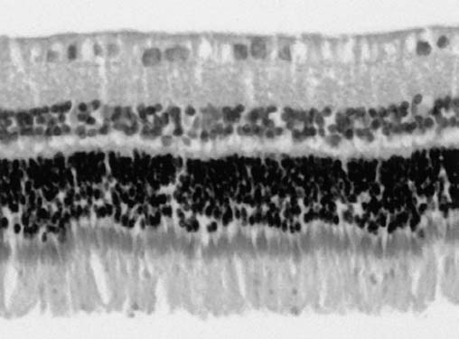
Fig. 1 Section of a rabbit retina 8 weeks after injection of 100 getanercept into the vitreous. No signs of toxicity are found. Theretina shows normal tissue structures
Fig. 2 The b-wave amplitudes in ERG recordings. Both treated anduntreated eyes were measured 4 and 8 weeks after injection of100 g etanercept
The histology of both treated and control eyes afterintravitreal administration of 100 g etanercept were notdistinguishable and showed normal anatomy (Fig. 1). Nosigns of toxicity were found. Quantification of retinallayers demonstrated no difference in either group.
A decrease in ERG b-wave amplitude was observed in
Fig. 3a–c Etanercept in various tissues at 0, 2, 4, and 8 weeks after
both treated and control eyes 4 and 8 weeks after injection
injection (amount of protein in relative fluorescence units): a invitreous; b in retina; c in choroid
of 100 g etanercept. Comparison of treated and control
The vitreous showed no opacities or bands formation.
eyes showed no reduction of b-wave amplitude as an
Such signs of toxicity can be found, for example, with
effect of treatment (Fig. 2). VEP could be elicited before
intravitreal amphotericin B injections. With increasing
and after treatment.
doses (10–50 g), retinal ganglion cell loss and focalnecrosis of the nerve fiber layer became also apparent [4].
Daunomycin used for inhibiting proliferative vitreo-
retinopathy has been reported to be toxic at doses of5 g in the vitreous [6].
Etanercept was measured in vitreous, retina, and choroid
Fluorescence-labeled etanercept was injected in two
at 0, 2, 4, and 8 weeks after injection of 5 g and 50 g
doses (5 g and 50 g) intravitreally and the amount of
fluorescein-coupled etanercept into the vitreous. In the
the protein was determined in vitreous, retina, and choroid
vitreous, a peak value was measured directly after
at 0, 2, 4, and 8 weeks after injection. By using gel
injection which gradually declined to the 8th week after
filtration, only the amount of intact etanercept was
injection. In both the retina and the choroid, the highest
determined. Therefore, a decrease in biological activity
values were found 4 weeks after injection. But even 8
by decay of etanercept can be ruled out. In the vitreous,
weeks after injection, etanercept was still found in retina
the amount of etanercept gradually declined up to the 8th
and choroid (Fig. 3).
week after injection. In both the retina and the choroid, aslow accumulation was found with a peak at 4 weeks.
After 8 weeks, etanercept was still clearly detectable. The
pharmacokinetics of etanercept after intravitreal deliveryensure high concentrations of the drug in the target tissues
Experiments with etanercept in a rat model of diabetic
over several weeks and offer the chance of a successful
retinopathy demonstrated that the drug reduces leukocyte
therapy. The relatively high molecular size of etanercept
adhesion, blood–retina barrier breakdown and endothelial
results in slower clearance than, for example, with
injury [8]. Therefore, diabetic retinopathy may be a new
triamcinolone [14, 15]. After injection of 400 g triam-
therapeutic indication for this agent apart from its main
cinolone acetonide, the half-life as determined by HPLC
indication in rheumatoid arthritis. As the intravitreal
was 1.6 days [15]. Dexamethasone, another glucocorti-
administration would minimize systemic side effects and
coid, has a half-life of only 2.5 h in physiologic saline [1,
deliver the drug locally, the pharmacokinetics and safety
of this route of administration were investigated.
In summary, the results show that intravitreally deliv-
After intravitreal injection of high doses of etanercept
ered etanercept is safe and leads to high concentrations in
(100 g per eye), no signs of toxicity were found on
the retina and choroid over several weeks.
evaluation of data from clinical examination, histology,ERG, and VEP. Etanercept did not induce any patholog-
Acknowledgements Supported by the DFG (Jo 324 /4-1, Jo 324/6-
ical changes after an observation time of up to 8 weeks.
1), the ZMMK Kln (TV76), the Ernst und Berta Grimmke Stiftungand the Gertrud und Werner Mller Stiftung.
Function and structure were the same as in control eyes.
1. Agabeyoglu IT, Wagner JG, Kay DR
4. Cannon JP, Fiscella R, Pattharachayakul
7. Limb GA, Chignell AH, Green W,
(1980) A sensitive high-pressure liquid
S, Garey KW, De Alba F, Piscitelli S,
LeRoy F, Dumonde DC (1996) Distri-
chromatographic method for the deter-
Edward DP, Danziger LH (2003) Com-
bution of TNF alpha and its reactive
mination of prednisone, prednisolone
parative toxicity and concentrations of
vascular adhesion molecules in fibro-
and hydrocortisone in plasma. Res
intravitreal amphotericin B formulations
vascular membranes of proliferative
Commun Chem Pathol Pharmacol
in a rabbit model. Invest Ophthalmol
diabetic retinopathy. Br J Ophthalmol
Vis Sci 44:2112–2117
2. Antoni C, Braun J (2002) Side effects of
5. Joussen AM, Murata T, Tsujikawa A,
8. Koizumi K, Poulaki V, Doehmen S,
anti-TNF therapy: current knowledge.
Kirchhof B, Bursell SE, Adamis AP
Welsandt G, Radetzky S, Lappas A,
Clin Exp Rheumatol 20:152–157
(2001) Leukocyte-mediated endothelial
Kociok N, Kirchhof B, Joussen AM.
3. Barouch FC, Miyamoto K, Allport JR,
cell injury and death in the diabetic
(2003) Contribution of TNF-alpha to
Fujita K, Bursell SE, Aiello LP,
retina. Am J Pathol 158:147–152
leukocyte adhesion, vascular leakage,
Luscinskas FW, Adamis AP (2000)
6. Hui YN, Liang HC, Cai YS, Kirchhof
and apoptotic cell death in endotoxin-
Integrin-mediated neutrophil adhesion
B, Heimann K (1993) Corticosteroids
induced uveitis in vivo. Invest Oph-
and retinal leukostasis in diabetes.
and daunomycin in the prevention of
thalmol Vis Sci 44:2184–2191
Invest Ophthalmol Vis Sci 41:1153–
experimental proliferative vitreo-
retinopathy induced by macrophages.
Graefes Arch Clin Exp Ophthalmol231:109–114
9. Limb GA, Soomro H, Janikoun S,
12. McLeod DS, Lefer DJ, Merges C, Lutty
16. Schrder S, Palinski W, Schmid-
Hollifield RD, Shilling J (1999) Evi-
GA (1995) Enhanced expression of
Schnbein GW (1991) Activated
dence for control of tumour necrosis
intracellular adhesion molecule-1 and
monocytes and granulocytes, capillary
factor-alpha (TNF-alpha) activity by
P-selectin in the diabetic human retina
nonperfusion, and neovascularization in
TNF receptors in patients with prolif-
and choroid. Am J Pathol 147:642–653
diabetic retinopathy. Am J Pathol
erative diabetic retinopathy. Clin Exp
13. Miyamoto K, Khosrof S, Bursell SE,
Immunol 115:409–414
Rohan R, Murata T, Clermont A, Aiello
17. Spranger J, Meyer-Schwickerath R,
10. Limb GA, Webster L, Soomro H,
LP, Ogura Y, Adamis AP (1999) Pre-
Klein M, Schatz H, Pfeiffer A (1995)
Janikoun S, Shilling J (1999) Platelet
vention of leukostasis and vascular
[TNF-alpha level in the vitreous body.
expression of tumour necrosis factor-
leakage in streptozotocin-induced dia-
Increase in neovascular eye diseases
alpha (TNF-alpha), TNF receptors and
betic retinopathy via intercellular ad-
and proliferative diabetic retinopathy].
intercellular adhesion molecule-1
hesion molecule-1 inhibition. Proc Natl
Med Klin 90:134–137
(ICAM-1) in patients with proliferative
Acad Sci USA 96:10836–10841
18. Tano Y, Sugita G, Abrams G,
diabetic retinopathy. Clin Exp Immunol
14. Schindler RH, Chandler D, Thresher R,
Machemer R (1980) Inhibition of in-
Machemer R (1982) The clearance of
traocular proliferations with intravitreal
11. Lu M, Perez VL, Ma N, Miyamoto K,
intravitreal triamcinolone acetonide.
corticosteroids. Am J Ophthalmol
Peng HB, Liao JK, Adamis AP (1999)
Am J Ophthalmol 93:415–417
VEGF increases retinal vascular ICAM-
15. Scholes GN, O'Brien WJ, Abrams GW,
1 expression in vivo. Invest Ophthalmol
Kubicek MF (1985) Clearance of tri-
Vis Sci 40:1808–1812
amcinolone from vitreous. Arch Oph-thalmol 103:1567–1569
Source: http://www.kalbacher.uni-tuebingen.de/pdf/2004/hk20044.pdf
Graefes Arch Clin Exp OphthalmolDOI 10.1007/s00417-012-2226-y Visual outcomes and complications following posterioriris-claw aphakic intraocular lens implantation combinedwith penetrating keratoplasty Johannes Gonnermann & Necip Torun &Matthias K. J. Klamann & Anna-Karina B. Maier &Christoph v. Sonnleithner & Antonia M. Joussen &Peter W. Rieck & Eckart Bertelmann Received: 21 August 2012 / Revised: 31 October 2012 / Accepted: 21 November 2012
Peninsula Health - Community Dental Program Dental Management of Presentation based upon DHSV Clinical Practice Guidelines Presented by Dr Fayed Azouz Clinical Head Peninsula Health – Community Dental Program 1) To inform the Peninsula Health Community Dental clinical staff on the Current DHSV Clinical Practice Guidelines for dental care management of the pregnant woman.