Le0409_00-00_reyhanian
I xxx _ xxx
The use of the Erbium Yttrium
Aluminium Garnet (2940nm) in a
Laser-Assisted Implant Therapy
How far we can push the envelope?
author_Dr Avi Reyhanian
fantasy? Does the new technology completely replace
The array of available clinical applications for laser
the conventional methods and if not, at which point
assisted dentistry is growing rapidly, with the greater
do we lay the laser's hand piece down and re-employ
number of applications being for oral surgery. Er: YAG
the "old" tools and conventional ways?
laser wavelength is considered to be extremely safe,
The article will exhibit, beyond any doubts, that
and is the dominant wavelength in dentistry today . Er:
Er:YAG laser is very valuable tools and shows promise
YAG is one of the most suitable wavelengths for bone
and safe as an effective new technical modality for im-
applications. The presentation will demonstrate the
plant therapy.
use of the Er:YAG laser in the world of implantology,
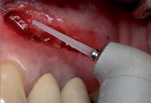
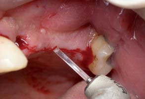
Fig. 1_intrasulcular incision
and the advantages vs. conventional treatment meth-
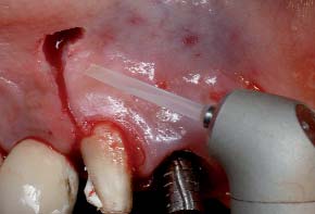
Fig. 2_Midcrestal incision
ods. The purpose in this paper is to put some order into
Osseo-integration dental implants have become a
Fig. 3_vertical incision for realese
the chaotic information surrounding the subject and
routinely recommended procedure in the clinical
Fig. 4_Semi lunar incision
to provide some answers to the most common and fre-
practice of dentistry 1-4, and have been utilized as a
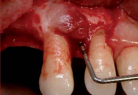
Fig. 5_Granulation tissue
quent questions we often meet: How far we can go
successful treatment modality over three decades,
Fig. 6_The erbium to ablate the gran-
with this technology? Is it just a marketing tool or
with a reported success rate of greater than 90% 5-7,8.
proven therapy? Where is the line between reality and
The predictability and success of dental implants has
xxx _ xxx I
secured their place as a standard treatment modality.
cooling: Extremely low thermal damage to surround-
Fig. 7_The site image immediate
This technique using the Er:YAG laser presents several
ing tissue. Can be used in contact or noncontact mode,
advantages vs. conventional treatment methods, and
efficient ablation effect for both soft and hard tissue.
Fig. 8_Marking the location of the
there are minimal post-operative complications cou-
Delivery system- articulated arm (Fotona), hollow
pled with a high rate of success. The Erbium :YAG is one
wave guide (Lumenis) , fiber optic ( Biolase, Doctor
of the most suitable wavelength for bone applications.
smile, Hoya combio,) or no delivery system (Lite-
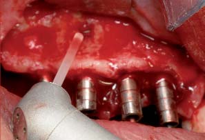
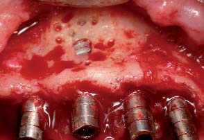
Fig. 10_The erbium for decortication
The Erbium's energy is highly absorbed in the water
Touch,Syneron medical ltd)
Fig. 11_Decotrication
component of dental tissue and provides efficient ab-
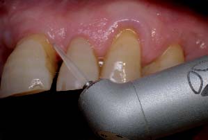
Fig. 12_Recontouring of the soft tis-
lation without the risk of significant thermal dam-
What Can We Actually Do Today ,2009, with the
sue around the implants
age.13 The Er:YAG laser is a thermal laser wavelength
Er:YAG Laser,In the World of Implantology
which is located in the infrared zone of the electro-
1- Incision- Midcrestal, Vertical , Intrasulculal
magnetic spectrum, considered to be extremely safe.
Most of the procedures performed at my practice are
2- Removal of granulation tissue. (Fig. 5, 6, 7)
in the realm of surgery and they are all performed with
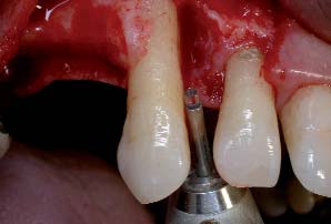
3-Marking the location and direction of the im-
the Er:YAG laser: Today we can observe an inordinate
plant – Only lase at the cortical bone. (Fig. 8)
lack clarity concerning the clinical uses of the laser in
4-Decortication and GBR technique. (Fig. 9, 10, 11)
the field of implantology, and with many doctors in the
5-Reshaping of the soft tissue (flap) around the im-
field reality does indeed mix with fantasy.
plants. (Fig. 12, 13, 14)6-Uncovering of submerged implants. (Fig. 15, 16)
What do we know about the Er:YAG laser- 2940 nm
7-Flapless implants.(Fig. 17,18,19,20)
The Erbium Yttrium Aluminium Garnet laser has a
8-Open sinus lift- Opening the window.
wavelength of 2940 nanometres' and emits as a free-
running pulsed train of photons in the mid infra-red
9-Periimplantitis treatment. (Fig. 23, 24, 25)
portion of the electromagnetic spectrum. Got FDA ap-
10-Implant failure treatment. (Fig. 26, 27)
proval for hard tissue in 1997. This laser is a thermal
11-Immediate implants in infected sites.
laser: interaction between the laser beam and the tar-
(Fig. 28, 29, 30, 31)
get tissue create heat. Successive laser pulses are 100-200 microseconds in width. The prime chromophore
1-The Erbium laser can make an incision for flap
of this laser wavelength is water, which makes it ap-
lifting 9-13, such as a crestal incision, or an intrasul-
propriate for ablating both hard and soft oral target
cular or vertical release incision. The laser produces a
tissue. Incident laser energy is absorbed by the chro-
wet incision (some bleeding) as opposed to the dry in-
mophore, converted into thermal energy which results
cision (no bleeding) that is produced by the CO2 laser.
in expansive vaporisation by micro explosions. Such
The tip of choice is chisel tip or 200 micron tip, contact
action causes a dislocation of the tissue structure and
mode, set power 300mj/25pps (7.5 watt.) When per-
ablation; often this is accompanied by an audible
forming the vertical incision, not to push the end of the
"popping" sound. Established high safety in clinical
sapphire tip into the soft tissue but rather to gently
use. The erbium laser presents availability of water
stroke the tissue with the tip. The doctor should lase
I xxx _ xxx
Fig. 13_immediate post op
the soft tissue until he feels the contact with the bone.
compared to the conventional osteotomies, and re-
Fig. 14_final result
2 Vaporization of granulation tissue 13,15 (if any ex-
sults of a study 20 indicated that in the rat model the
Fig. 15_The erbium to uncover the
ists) after raising a flap is efficient with the Erbium
lased bone prepared implant sites vs. conventional
submerged implant
laser, with a lower risk of overheating the bone
bone preparation sites developed a statistically higher
Fig. 16_The uncovered implant
13,16,17 than those posed by the current diode or CO2
percentage of bone-to-implant contact. 4 One of the
Fig. 17_The erbium to lase the soft
lasers. Chosen laser is Er:YAG (LiteTouch, Syneron
golden rules for GBR-Guided Bone Regeneration is to
medical ltd). Tip of choice is 1300 micron, noncontact
Provide a good blood supply – Cortical stimulation: To
Fig. 18_The soft tissue after lasing
mode (Distance between end of the tip and target tis-
perforate the cortical bone to get bleeding which bring
sue = 1.5 mm) set power 400mj/ 17pps = ( 6.8 watt). It
with him mesenchimal cells, they transform to os-
is important to vaporize the granulation tissue before
teoblasts, they transform to osteocits which are re-
drilling for the implant because one of the reasons for
sponsible for bone formation. Decortication is per-
IPL (implant peri-apical lesion) is insertion of soft tis-
formed with the erbium laser (LiteTouch, Syneron
sue into the site preparation for the implant. 3 Using
medical ltd). The tip of choice is 1300 micron, Set
the Erbium laser in non-contact mode (1.5-2 mm from
power 300mj/25pps (6 watt). Noncontact mode. No
the target tissue), the future location and angle of the
rotary tool vibrations: reducing patient discomfort
implant is outlined; and the laser is used only on the
and enhancing the surgical site. Less stressful oral
cortical bone. Set power: 300mj/20 pps (6 watt). As an
therapy with enhanced outcomes.21,15 5 Dental im-
important point of clarification, the laser does not re-
plant must form and maintain integration not only
place the pilot drill; it is used to create a "pilot hole" for
with bone but also with connective tissue and epithe-
the drill. The entire length of the implant is not lased
lium- The Junctional epithelial attachment is an im-
with the laser. This technology doesn't give, yet, the an-
portant component of the protective permucosal soft
swer to the question how to lase all the entire length
tissue seal and may even limit the apical spread of mar-
for the implant with out the use of rotary instruments.
ginal inflammatory that can lead to bone loss or im-
Although there are some dental lasers users with a lot
plant failure. If there is enough keratinized tissue, be-
of experience who claim that they lase all the entire
fore closing the flap it is recommended to reshaping
length of the implant with the erbium .This statement
the soft tissue (flap) around the implants in order to
raze 2 main concerns: The first one is how to control
get a better sealing. Tip of choice 200 micron, contact
the depth and the second one, Which is the the fun-
mode, Set power 250mj/30pps (7.5 watt). 6 Implant
damental concern in any bone surgical procedure, is
exposure can be done with the Er:YAG, with the Diode
how to limit thermal rise to within 470C 18 in order to
and with the co2 laser or other wavelengths. The big
avoid damage to cellular components of bone metab-
advantage of the erbium in this procedure versus the
olism and delayed healing 19. Further clinical and ba-
others is less zone of thermal necrosis because of the
sic investigations are require to establish the clinical
water spray cooling. The benefits of laser use over
effectiveness and safety of the Er:YAG laser in implant
scalpel include precision, incisional hemostasis and
site preparation. We are not there yet although Er:YAG
immediate post-operative protection through a tena-
laser can promote the growth of new bone around
cious coagulum surface 22, 23 Local anesthetic may
placed titanium implants and better osseointegration
or may not be used, depending on patient and opera-
xxx _ xxx I
tor preference. A small cone of tissue is removed until
from the attachment of the flap to minimize the risk of
Fig. 19_drilling for the implant
near-contact with the screw is made. Tip of choice can
an air embolism. Antrostomy was performed with the
Fig. 20_Insertion of the implant
be the 200 or 400 or 600 or 800 or 1000 micron. The set
Erbium laser to create a rectangular shape26. The
Fig. 21_The erbium to open the lat-
power is around 6 watt. 7 In those crests in which the
height of this window should not exceed the width of
width is more than 7 mm, it is possible to insert the im-
the sinus. After lasing all four flanks of the window, the
Fig. 22_The lateral window per-
plant without razing a flap. With the erbium laser a
bone was gently removed or pushed inside the sinus,
formed with the erbium
small cone of tissue is removed with the diameter of 6
taking care not to damage the Schneiderian mem-
Fig. 23_The erbium to ablate the
mm. (Tip sapphire 800 mic, contact mode, set power
brane 27. The sinus membrane was then lifted gently
granulation tissue
300/25pps= 7.5 watt). With the 1300 mic sapphire tip,
from the bony floor .A space was created after the si-
Fig. 24_Detoxification of the infected
in non-contact mode (1.5-2 mm from the target tis-
nus membrane has been elevated, which was then
sue), the future location and angle of the implant is
grafted with different material.
outlined; and the laser is used only on the cortical
Experience has shown that the laser is safer to sur-
bone. Set power 300mj/25pps (7.5 watt). In the mo-
rounding tissue than rotating instruments 27, 28, 29
ment the location of the implant is marked it is rec-
particularly when it comes to the risk of perforating
ommended to reemploy the rotary instrument. 8 An
the Schneiderian membrane. When adhering to the
Er: YAG laser has been determined as the most suitable
recommended operating parameters and tools (en-
platform for this procedure. The Erbium family of
ergy, tips, hand piece configuration and mode of beam
lasers is the only one which delivers a tissue-cooling
application - contact/non-contact). 9 Periimplantitis
water spray together with the laser beam, an ex-
is an inflammatory reaction that is associated with the
tremely important feature when lasing bone tissue
presence of a sub-marginal biofilm, with advanced
10,24 25. The tip of choice was a 1300 micron sapphire,
breakdown of soft and hard tissue surrounding the en-
using non-contact mode. The distance between the
dosseous implant: loss of the bony support of the im-
end of the tip and the target tissue should be 9mm. The
energy used for this procedure was 200 milliJoules / 15
Therapeutic objectives focus on correcting techni-
PPS, (3W average power). The headpiece should be al-
cal defects by means of surgery and decontamination
ways in constant motion very close attention paid, be-
techniques: REJUVENATING. Removing mobile im-
cause the bone of the lateral window is usually very
plants is recommended.
thin. The Er: YAG laser does not provide good hemo-
Therapeutic and surgical approaches in the con-
stasis, both because of its short interaction time and
ventional system include:
its shallow depth of penetration. The moment a littlebleeding appears, or the Schneiderian membrane is
1. Systemic administration of antibiotics
seen, the laser beam should be moved forward. The
2. Removal of supra-gingival bacterial plaque
closer the beam comes to the Schneiderian membrane,
3. Removal of granulation tissue with plastic
the greater the distance should be between the tip and
the target tissue (the energy is controlled by the dis-
4. Detoxification of the exposed surface 31
tance between the end of the tip and target tissue).
_Mechanical brushing
Pressurized air in the water spray was directed away
_Air powder abrasive
I xxx _ xxx
11-Insertion of implants in infected sites presents
a lot of problems and some times require 2 stages of
_Topical tetracycline application
surgery. In conventional methods there is no tool in
_Low intensity UV-radiation
which in the same time provides decontamination of
5. Removal of peri-implant
the target tissue: By lasing with the erbium it is possi-
ble to gain decontamination of the infected site, which
6. Regeneration of peri-implant
enables the doctor to save chair time and, in some of
hard tissue (GBR)
the cases, to avoid the second stage of implant surgery.
Fig. 25_The exposed screw after
7. Plaque control
Fig. 26_Two mobile implants engage
In addition to conventional treatment modalities,
Er:YAG laser is an essential and indispensable tool
the use of the Er: YAG laser has been increasingly pro-
for implant surgery today.
Fig. 27_The erbium to ablate and de-
moted for the treatment of peri-implantitis:
This wavelength shows promise and safe as an ef-
contaminate the site
fective new technical modality for implant therapy
Fig. 28_At presentation
_The Erbium laser can make an incision for flap lift-
when adhering to the recommended operating pa-
Fig. 29_The erbium to ablate granu-
rameters and tools (Energy, tips, hand piece configu-
lation tissue and decontamination
_Vaporization of granulation tissue
ration and mode of beam application - contact/non-
Fig. 30_Immediate implants
_Detoxification of the implant surface32 by lasing
contact). However, further clinical and basic investi-
Fig. 31_Immediate implants
directly on the implant's exposed screws, using a low-
gations are requiring for establishing the clinical ef-
energy setting, the target tissue is disinfected together
fectiveness and safety of the Er: YAG laser in implant
with the implant surface15 without injuring their sur-
site preparation._
faces 33-35.
The Literature list can be requested from the editorial
_The laser is bactericidal 36, 37. The significant bac-
terial reduction in the implant surface and the periim-plant tissues during irradiation are the main reasonsfor the erbium laser application in the treatment of
Dr Avi Reyhanian
_Ablating the bone with the Er:YAG laser: remod-
Private Clinic ,Netanya, Israel
eling, shaping and ablating necrosis Bone 38,39,40,14
1 Shaar Haemek St. Netanya 42292, IsraelOffice: +972-9-8338825
10- In implants failure sites treatment the Er:YAG
Fax: +972-9-8339890
laser assist the doctor by the mean of incision to open
the flap, ablation of granulation tissue, ablation of
necrotic bone and decontamination of the site for GBR
Source: http://tomasfoustka.cz/wp-content/uploads/2013/05/Implantace-spomoc%C3%AD-laseru-%C4%8Dl%C3%A1nek.pdf
Comparative efficacy of haloperidol and olanzapine in patients of schizophrenia: a 6 month follow up trial Romika Dhar, BS Chavan, Ajeet Sidana Abstract : P resent study was carried out to compare the efficacy f olanzapine and haloperidol in patients of schizophrenia. A prospectiv randomized, open label studycarried out on patients attending the outpatient clinic or those admitted to inpatientservices. 40 cases of schizophrenia diagnosed as per ICD-10 were randomized toolanzapine and haloperidol group and were assessed at 3 months andafter 6 months of treatment using scales PANSS and ESRS. the study showed thatboth the drugs were equally efficacious in improving the psychopathology andhaloperidol led to increase in extrapyramidal side effects. Both olanzapine andhaloperidol are equally effective in causing clinical ment . In comparison,both the drugs do not have much difference in the cost of treatment.
AM P: SLTOPHO Acknowledgments The content of this booklet was researched and written by Dr. Janet McKeown (MD, CCFP, DipSportsMed), Cristina Sutter (Registered Sport Dietitian) and Susan Boegman (Registered Sport Dietitian) with input from Dr. Penny Miller (Clinical Pharmacologist), Dr. Susan Hollenberg (Family & Travel Medicine Physician), and Dr. Reka Gustafson (Medical Health Officer).


