Yloop.in
ISSN 2349 – 4425
www.americanij.com
The Protective Effect Of Some Natural
Antioxidants Against Azithromycin Induced
Testicular Dysfunction In Rats
El-Dakak, Abeer M. N. H.Ph.D
Special Food and Nutrition Dept., Food Technology Research Institute, Agric. Res. Center, Giza, Egypt
Email address:
[email protected]
Abstract:
Objective: To investigate whethervitamin E, selenium, β-carotene, anthocyanin and green tea extract have a
possible protective effect against azithromycin (AZ)-induced testicular dysfunction.
Design: Experimental study.
Setting:Research Institute of Ophthalmology, Giza, Egypt.
Patient(s):3 -week-old weanling male albino rats (n = 54).
Intervention(s):AZ was administered to rats at dose of 20mg/kgbw orally for 3 days every two weeks.
Vitamin E, β-carotene pigment,anthocyanin pigment, green tea extract, Se as sodium selenite and their
mixture were administered with AZ by gavage daily for 4 months.
Main Outcome Measure(s):Body and reproductive organ weights, spermatozoal activity, fructose contents,
testosterone (T), interstitial cell-stimulating hormone (ICSH), lipid peroxidation and, antioxidant enzyme
activities were investigated.Histopathological changes in reproductive organs examined.
Result(s):Significant decreases in serum T and ICSH,reproductive organ weights, spermatozoal activity and
fructose content were recorded due to AZ administration, whereas these parameters increased the
antioxidants-treated groups. A significant decrease in Testicular MDA level and marked increases in GSH-
Px, SOD, and CAT activities were observed in rats treated with different antioxidants. However, the most
positive treatment was Se-treatment and the mixture of vitamin E with β-carotene pigment. AZ-treatment led
to adverse histopathologic changes in the testes, epididymis cuda, seminal vesicle and prostate, whereas
normalhistopathological imageof the studied reproductive parts were observed in groups treated with Se,
Vitamin E and β-carotene.
Conclusion(s):Antioxidants have a protective effect against testicular toxicity caused by AZ. This protective
effect of the antioxidants seems to be closely involved with the suppressing of oxidative stress.
Key words: Azithromycin; Antioxidants; Oxidative stress; spermatozoal activity; Testes.
Volume 2 2015 Issue 1 DEC-JAN AIJCSR
ISSN 2349 – 4425
www.americanij.com
occurring prior to in-vitro fertilization (IVF) treatment, or
1. INTRODUCTION
when high concentrations of leukocytes are present in the
Infertility or sterility is defined as the total inability to
semen of these patients, irrespective of microbial evidence
reproduce
(1); it has been estimated that 7.2% of women and
of infection. Patients on a course of antibiotics often
their husbands in Egypt 2012 are infertile according to
demonstrate below-average semen parameters. While in
WHO, 2013
(2). Infertility has an element of time in its
some instances this may be caused by the infection itself, it
definition;it can be stated as failure to conceive after one
is likely that the antibiotics have a direct effect on sperm
year of regular unprotected intercourse with the same
function
(10). Studies also have shown that antibiotics
partner
(3). Infertility can be classified into primary and
e.g.,Cotrimoxazole, dicloxacillin, erythromycin, lincomycin,
secondary, A male without biological offspring can be said
neomycin, nitrofurantoin, quinolones, tetracycline, and
to have ―primary infertility,‖ whereas a male who is unable
tylosin;considerably impair sperm motility characteristics
to impregnate his partner but who already has biological
(8,11) and functional integrity of mature sperm
(12).
children is referred to as having ―secondary infertility‖
(4).
Furthermore, perfloxacin produced toxic effect on testicular
For healthy young couples, the probability of getting
function in animals
(13).
pregnancy per a reproductive cycle is about 20% to 25%.
Their cumulative probabilities of conception are 60% within
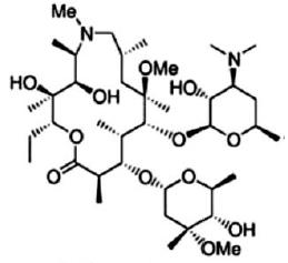
homoerythromycin,
Fig.(1), an erythromycin semisynthetic
the first 6 months, 84% within the first year, and 92% within
derivative is formed by inserting a methyl-substituted
the second year of regular fertility-focused sexual activity
nitrogen in place of the carbonyl group at the 9a position of
the aglycone ring. The resulting dibasic 15-membered ring
Several studies have suggested that human semen
macrolide derivative is more appropriately referred to as an
quality and fecundity have been declining during the past
‗‗azalide.‘‘ This structural modification gives azithromycin
decades
(6). Approximately 10-15% of couples demonstrates
several distinct advantages over erythromycin: makes the
primary infertility and of these a male factor is identified in
compound more stable in acid, significantly increases the
approximately 50% of cases
(7).Many extrinsic and
environmental factor including the increased use of
gastrointestinal tolerability, oral bioavailability and tissue
antibiotics have been implicated as potential causes of male
penetration, and results in increased activity against gram-
negative organisms when compared with erythromycin
(14).
inflammatory diseases (PID) and sexually transmitted
Its extensive coverage against all the major respiratory
infections (STI).In addition personal habits that considered
pathogens, excellent safety profiles and desirable tissue
risk factors for infertility, i.e., excess alcohol intake,
distribution into the infection sites make azithromycin a
cigarette smoking and other forms of drug abuse. Therefore,
good choice for the treatment of respiratory tract infections
Onyije (9) revealed that drugs such as antibiotics, alcohol,
(15).Azithromycin is frequently prescribed for the treatment
tobacco products, illicit drugs and certain medications can
of middle ear and upper respiratory tract infections,
limit the chances of men having children in the present time,
bronchitis, and community-acquired pneumonia
(16).In
or even far into the future.
recent years, it has been used primarily to prevent bacterial
Antibiotics such as amoxicillin and tetracycline are
infections in infants and those with weaker
commonly prescribed for a multitude of conditions
(10), in
most cases these antibiotics are used in treating infections
Macrolide antibacterial agents including azithromycin are
that are unrelated to infertility. In addition, some patients
lipophilic and are extensively distributed in body fluids and
requiring assisted conception occasionally show evidence of
tissues. They exert their antibacterial effects by reversibly
infection on the male reproductive tract. The antibiotics co-
binding to the 50s subunit of the bacterial ribosome. This
trimoxazole and erythromycin are routinely used by
interaction inhibits RNA-dependent protein synthesis by
urologists and fertility specialists to treat bacterial infections
preventing transpeptidation and translocation reactions
(18).
Volume 2 2015 Issue 1 DEC-JAN AIJCSR
ISSN 2349 – 4425
www.americanij.com
and produce infertility by two key mechanisms. First, ROS
damage the sperm membrane, which in turn reduces sperm's
motility and ability to fuse with the oocyte. Secondly, ROS
directly damage sperm DNA,compromising the paternal
genomic contribution to the embryo. Despite the common
association between compromised sperm quality and
oxidative damage, men are rarely screened for oxidative
stress nor treated for this condition(23).
In more details, free radicals have the ability to
directly damage sperm DNA by attacking the purine and
pyrimidine bases and the deoxyribosebackbone. Normally,
sperm DNA is tightly packaged by protaminesprotecting it
from free radical attack. However, infertile men often
exhibit deficient protamination, leaving the spermDNA
particularly vulnerable to ROS attack (24). Alternatively,
free radicals can initiate apoptosis within the sperm, leading
to caspase-mediated enzymatic degradation of the DNA
Fig. 1. Azithromycin
Certain macrolide antibiotics have been documented to be
hepatotoxic in animals (19) and human subjects in clinical
highunsaturated fatty acids and are sensitive to oxygen
studies (20). Macrolide antibiotics are also known to alter
induced damage mediated by lipid peroxidation. The
the physiological redox homeostasis leading to oxidative
excessive generation of ROS by abnormal spermatozoa and
stress and lipid peroxidation(21).
by contaminating leukocytes that has been identified as one
Cells require oxygen for certain metabolic processes
of the few etiologies for male infertility (26).
including the metabolism of xenobiotic and in the process of
Actually, a balance between the benefits and risks from
certain destructive species referred to as reactive oxygen
reactive oxygen species and antioxidants appears to be
species (ROS) are generated (22). The cell protects itself
necessary for survival and normal functioning of
from these ROSs, by the actions of certain low molecular
spermatozoa (27). Increased production of lipid peroxide is
weight substances such as glutathione, vitamin A, E, and C
associated with decrease in Zinc as antioxidants .An
(the non enzymic antioxidants) and enzymic antioxidants
imbalance in this harmony may be responsible for the
such as superoxide dismutase (SOD), catalase (CAT) etc.
inactivation of hyaluronidase activity thereby leading to
Therefore, the objective of this study was to investigate the
severity of oligozoospermia(28).Yadavaet al.,(29) observed
effect of azithromycin as antibiotic on the markers of
that decrease in total antioxidant power correlates with
testicular function like a sperm quality, spermatogenic cell
abnormal sperm membrane integrity by ROS and increased
density, testosterone level, antioxidant activity and lipid
nitric oxide concentration. This suggests that imbalance
peroxidation in male rats.
between antioxidants and reactive nitrogen species results in
Oxidative stress (OS) occurs when the production
sperm membrane integrity.
of potentially destructive reactive oxygen species (ROS)
It has been proposed that bactericidal antibiotics
exceeds the bodies own natural antioxidant defenses,
can induce cellular death through a common oxidative
resulting in cellular damage. OS is a common pathology
damage mechanism that relies on the production of
seen in approximately half of all infertile men. ROS, defined
ROS.Through their various primary targets, antibiotics can
as including oxygen ions, free radicals and peroxides are
activate cellular respiration, which leads to the formation of
generated by sperm and seminal leukocytes within semen
superoxide and the release of iron from iron-sulfur clusters.
Volume 2 2015 Issue 1 DEC-JAN AIJCSR
ISSN 2349 – 4425
www.americanij.com
Free iron then activates a chemical reaction, i.e., the Fenton
allergies and co-administrated drugs, is important in order to
reaction) to produce ROS in the form of hydroxyl radicals
minimize the risk of adverse reactions (34). The side effects
(OH•). These radicals can cause cellular death by damaging
of antibiotics on spermatogenesis are seldom discussed in
proteins, lipids, and DNA, or can cause mutations leading to
the literature. The increaseincidence of sterility in man along
the development of antibiotic resistance(30).Nguyen,(31)
with increase in number of new therapeutic drugs
found that mutant bacteria deficient in the stringent response
administrated to patients has made experimental work on
exhibited tolerance to a wide range of antibiotics including
antibiotics in male reproduction important since these
ofloxacin, meropenem, colistin, and gentamicin by
medication found to adversely affect male reproductive
increasing antioxidant enzyme production and blocking the
functions (35).
production of pro-oxidant molecules, thus reducing toxic
However, the mechanism(s) by which antibiotics
OH•. These mutant bacteria also were more susceptible to
exert their antifertility effect in the male are yet to be fully
ofloxacin in a mouse infection model. Moreover, Goswami,
elucidated andseveral studies have demonstrated the
et al., (32) studied the effect of dietary and cellular
antifertility effects of free radicals and numerous
antioxidants on antibacterial effect of commonly used
mechanisms of action have been proposed (9, 11,13).Hence,
antibiotics. Dietary supplements such as vitamin C
previous investigationssuggested that OS may be an
(Ascorbic acid) and E (-tocopherol), having antioxidant
important mediator of testicular injury. The present study
properties, are prescribed nowdays by the physicians along
was therefore designed to investigate the role of some
with antibiotics during the course of treatment of infection.
e.g., vitamin E, β-carotene,
Besides antioxidants such as N-acetylcysteine (NAC) are
anthocyanins, polyphenols in green tea extract and selenium
used as auxiliary medication in certain pathological
on azithromycin (antibiotic) induced alterations which
conditions along with the antibiotic therapy (NAC is used as
massively used during early age cycles and its effect on
a mucolytic agent in combination with clinically relevant
male reproductive functions.
antibiotics for treatment of lower respiratory tract infection).
Therefore it is important to understand the effects of
2. Materials and Methods
antioxidants on theantibacterial action of commonly used
2.1. Materials
antibiotics. Metabolic action of eachantibiotic should be
2.1.1.Lyophilized green tea extract:Ten volumes of
taken in account when applied.
boiling water were added to tea (10v/ wt) and allowed
Antioxidant mediated protection against ciprofloxacin could
standing for 30 min at room temperature and the extracted
be through scavenging of ROS generated in the presence of
was filtered and lyophilized (36). The lyophilized green tea
antibiotic. Consequently effect of mutations in oxidative
extract used in this study contained 21.87 g catechins /100g
stress defense genes viz. superoxide dismutases (sodA,
green tea) was measured by HPLC.
2.1.2.Lyophilized anthocyanins pigments from red grape
hydroperoxidereductase
marc: The lyophilized anthocyanin pigment was prepared
susceptibility of MG1655 was studied. These genes encode
according to the method reported by Revilla, et al., (37) and
enzymatic defense system against ROS, regulating their
modified by El-Dakak (38). It was composed mainly of
intracellular steady-state level(32).
delphinidin 3-O-glucoside (Df-Gl), cyanidin 3-O-glucoside
In another case, oral antimicrobial agents belonging
(Cy-Gl), petunidin 3-O- glucoside (Pt-Gl), peonidin 3-O-
to beta-lactams, quinolones, macrolides, tetracycline and the
glucoside (Pn-Gl), malvidin 3-O-glucoside (Mv-Gl),
trimethoprim-sulfamethaxazole combination are among the
malvidin 3-O- acetylglucoside (Mv-Gl-Ac) and malvidin 3-
most prescribed classes of drugs in medical practice(33).
O-p-coumarylglucoside (Mv-Gl-Cou) as measured by
Again, knowledge of the potential side effects considered in
HPLC. Total amount equivalent to 89.84g/ 100g lyophilized
the light of various patient associated factors such as genetic
anthocyanin pigment.
make-up, renal and liver function, underlying disease, drug
Volume 2 2015 Issue 1 DEC-JAN AIJCSR
ISSN 2349 – 4425
www.americanij.com
2.1.3.Lyophilized β-carotene pigment from Carrot pulp
Rats were observed daily for the appearance of any
waste:HPLC analysis indicated that β-carotene was found to
symptoms of discomfort that might be related to studied
be as 83.8% in the lyophilized β-carotene pigment; which
treatments. Body weight (BW) of the rats was recorded daily
was prepared according to the method reported by Chen
before administration of studied additives. At the end of
&Tang (39) andmodified byEl-Dakak (38).
experiment period, the percentage of weight gain was
2.1.4. Drugs:Azithromycin (Zithromax® 250 mg and 500
expressed [(final weight – beginning weight) / beginning
mg tablets) was a product of Pfizer, New York.
2.2.3. Weight of reproductive organs
2.1.5. Animals:MaleWister strain albino rats weighing45 g
± 2 (3 weeks age) obtained from Research Institute of
At the end of the experimental period, rats were weighted
Ophthalmology, Giza, Egypt, were used for the study. Rat
and killed by diethyl ether. Testes, epididymis, seminal
cubes were fed on laboratory chow and tap water ad
vesicles and prostate were cut off, washed in ice-cooled
libitum4 months.The animals were housed in separate
saline solution0.15M KCl to remove blood and weighed.
stainless steel cages raised in a well-ventilated room with
Organ weights were recorded after autopsy and represented
12-h light/dark cycle.
as weight per 100 g body weight (42).
Right hemiprostate and seminal vesicle with the adherent
2.2. Methods
coagulating gland were removed and stored at –20°C until
2.2.1.Experimental design:All animal rights have been
fructose content was determined (43).
considered during all periods of the experiments according
2.2.4.Spermatozoal activity
to the Animal Care and Use Committee (IACUC)
The Resazurin Reduction Test (RRT) was applied for
guidelines. Fifty four male albino rats (9/group of six each)
determining spermatozoal activity (44). RRT depends on the
wererandomlyallotted into experimental groups as follows.
ability of metabolically active spermatozoa to reduce the
Group1 (negative control) were administered with distilled
resazurin dye (blue), with maximum absorption at 615 nm
water (vehicle for the drug). The other rats treated with
(A615), to resorufin (pink) with a maximum absorption of
20mg/ kgbw of azithromycinby oral gavage once daily at an
580 nm (A580). The ratio of the optical densities of reduced
interval of 24 hr. between successive treatments,for 3 days
to oxidized form, i.e., 580 to 615 nm can be used to evaluate
every two weeks(which represents the therapeutic dose of
the various grades of semen sample. Semen samples were
the drug in humans),then divided into 8 groups; group II
centrifuged at 2500 g for 20 min. The seminal plasma
rats,which also served as the positive control.After
supernatant was applied for test according to manufacturer's
azithromycin administration, rats in groups III and IV were
constructions using kits purchased from Bio-diagnostic,
co-treated orally with Vitamin E (3IU /kg bw), β-carotene
(50mg/kg bw) in corn oil, per day; respectively.Ingroups V,
2.2.5. Biochemical assay
VI and VIIrats were co-treated orally with anthocyanin
At the end of experimental period, blood samples were
(71mg /kg bw), green tea (120mg/kg body weight), with
collected from the eye plexuses of animals by a fine
selenium (0.1 mg/kg bw) as sodium selenite in deionized
capillary glass tubes and placed immediately on ice. Blood
water a day, respectively.Lastly, groups VIII and VIIIIrats
serum samples were collected into dry clean centrifuge
were co-treated orally with anthocyanins (24mg /kg bw)+
tubes; the serum was separated after centrifugation for 10
green tea extract (40mg/kg bw) + Se (0.03mg sodium sel/kg
min at 3000 rpm (1500 xg) and kept at –20 ºC until analysis.
bw)in deionized water; Vitamin E (1.5 IU /kg bw), β-
Testosterone and interstitial cell-stimulating hormone
carotene (25mg/kg bw) in corn oil, per day; respectively(40,
(ICSH) concentrations were measured in triplicate by
41). The Antioxidants application was given throughout the
ELISA according to the producer's instructions (Gama
whole trial except time of drug administration.
2.2.2.Body weight gain (BWG)
2.2.6.Biochemical analysis of testis
Volume 2 2015 Issue 1 DEC-JAN AIJCSR
ISSN 2349 – 4425
www.americanij.com
The right testis homogenate was centrifuged at 800
testis and epididymis as compared with the control animals
rpm for 20 min at 4°C. The supernatant was used for
(P<0.05) (Table 1). Azithromycin also caused decrease in
biochemical analysis.
weights of seminal vesiclesand prostate gland when
2.2.6. 1.Protein
expressed as mg/ 100gm body weight(Table 1). Co-
The protein level in the testis homogenate was
administration of azithromycin with different treatments
assessed by the method described by Gornall&Bardawill
significantly prevented the decrease in the weights of the
(45)to express other biochemical parameters as per mg
prostategland. However, the remained accessory glands
proteinusing kits obtained from Randox Laboratories Ltd.,
weights significantly increased by different antioxidant. Co-
Antrim, United Kingdom.
treatment of selenium (0.1 mg/kg bw) recorded the highest
2.2.6. 2.Estimation of enzymatic antioxidants
value in the relative testicular weights and weight of seminal
The superoxide dismutase (SOD),catalase (CAT),
vesicles. However, co-treatment of anthocyanin (24mg /kg
and glutathione peroxidase (GSH-PX) in the testis were
bw)+ green tea(40mg/kg bw) + selenium (0.03mg/kg bw)
estimated according to (46- 48)respectively, using kits
appeared to be the highest value in the relative cauda
obtained from Biodiagnostic.
epididymis weight.It is to theorizethat is the weight of
2.2.6. 3.Lipid peroxidation
sexual accessory gland decrease following azithromycin
The Lipid peroxidation (Malondialdehyde,MDA)
administration may be explained by reduction of plasma
was estimated according to Metlzer et al (49), using kits
level of testicular androgens.
obtained from Biodiagnostic.
Table 1.Body weight gain and weight of reproductive
2.2.6.4. Fructose content
organs of male rats in four months of treatments.
Fructose content of accessory sex glands (testis,
Weight of organs (mg/ 100 g BW)
right hemiprostate and seminal vesicle with the adherent
Daily intake
weight gain
coagulating gland) wasdetermined using the procedure of
Mann (50) using kits purchased from Bio-diagnostic, Egypt.
2.6. Histopathological studies
Different sections of studied reproductive organs (left testis,
control,
basal diet)
caudaepididymes, seminal vesicles and prostate) were
prepared for histological examination (51).
2.7. Statistical analysis
(Control
treated with
All values are means ± SD obtained from eight animal
20mg kg-1bw
groups (six of each). Data were analyzed with SAS software
(SAS Institute, Cary, N.C.) using SAS analysis of
Vitamin E(3
variance(PROC ANOVA). Significant differences between
IU /kg bw)
means were determined by Duncan's multiple range test (P <
(50mg/kg
3. Results
3.1.Weight gain and reproductive organs weight
The body weight gain and relative testicular weights and
(120mg/kg
accessory glands weights are presented in Table 1. The body
weights of the treated rats did not show significant changes
Selenium (0.1
mg/kg bw)
(Table 1). In the group treated with azithromycin alone,
there was significant decrease in relative weights of the
Volume 2 2015 Issue 1 DEC-JAN AIJCSR
ISSN 2349 – 4425
www.americanij.com
(mg/kg BW)
[Anth (24mg
/kg bw)+ ,
(40mg/kg
(0.03mg/kg
control, basal diet)
[Vitamin E(
(Control 1.23±0.14g
0.74±0.18f
1.5 IU /kg
treated with 20mg kg-1bw
carotene
of azithromycin)
(25mg/kg
Abbreviations; t = testes, p = prostate, c = cauda epididymis, sv = seminal
Vitamin E(3 IU /kg bw)
4.89±0.09c
1.38±0.22e
vesicles. Values are means ± SD (n=6);within each classvalueswith
different superscripted letters in the same column are significantly different
(p < 0.05).
β-carotene(50mg/kg bw)
2.48±0.30e
3.2.Serum the testosterone (T) and interstitial cell-
1.69±0.12f
stimulating hormone (ICSH) concentrations
The levels of T and ICSH in rats treated with
azithromycin, Vitamin E, β-carotene, anthocyanin, green tea,
5.52±0.36b
2.43±0.2bc
and selenium are shown in Table 2. Azithromycin
extracted(120mg/kg bw)
administration significantly decreased serum T and ICSH
concentration (P < 0.05) compared to the control group. Co-
Selenium (0.1 mg/kg bw)
8.55±0.42a
3.68±0.30a
administration of Vit E, β-carotene, anthocyanin, green tea,
selenium, mixture of the (anthocyanin+ green tea +
Anthocyanin+ Green tea
3.43±0.36d
2.83±0.74b
selenium) and mixture of the (Vitamin E+ β-carotene)
extract +Se
increased (P <0.05) the testosterone levels by (276, 32), (91,
147), (31, 146), (325, 230), (559, 399), (164, 283) and (310,
[Anthocyanin (24mg /kg
373)respectively, relative to the azithromycin-treated
bw)+ , green tea extract
(40mg/kg
(0.03mg/kg bw)]
Table 2. Testosterone (T) and interstitial cell-stimulating
hormone (ICSH) level of secretion in blood serum of male
Vitamin E+ β-carotene
5.32±0.28b
3.49±0.20a
rats in four months of treatments.
[Vitamin E( 1.5 IU /kg
(25mg/kg bw)]
Values are means ± SD (n=6);within each classvalueswith
different superscripted letters in the same column are
significantly different (p < 0.05).
Daily intake
Volume 2 2015 Issue 1 DEC-JAN AIJCSR
ISSN 2349 – 4425
www.americanij.com
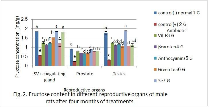
3.3.Fructose content and spermatozoal activity in the
reproductive organs
Fructose content of the four tested reproductive
organs were determined and represented in Fig. 2.
Azithromycin administration significantly decreased the
fructose content of prostate and seminal vesicle plus
coagulating gland in relation to the negative control group.
Clearly, all treatments applied in the present study led to
significant increase in the fructose level in the sexual studied
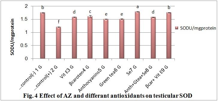
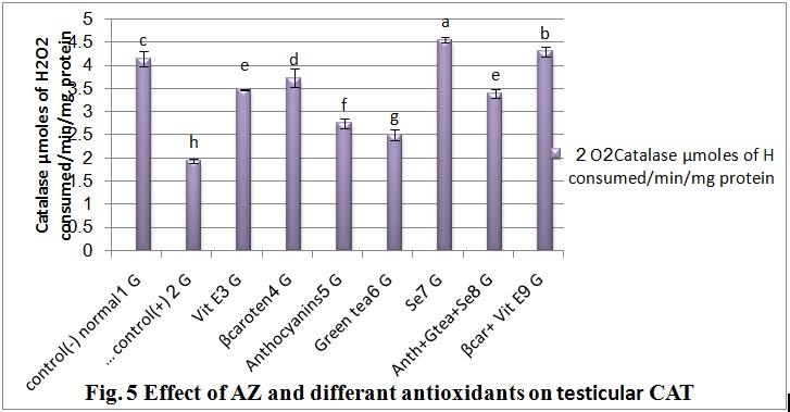
3.3.Antioxidant and testicular markers enzymes
organs. However, the severest treatment was selenium
In order to explore the possibility that azithromycin
treatment (G7).Although, the use of mixture of vitamin E
interferes with antioxidant defense system and thereby
with β-carotene (G9), especially seminal vesicle(Fig. 2).
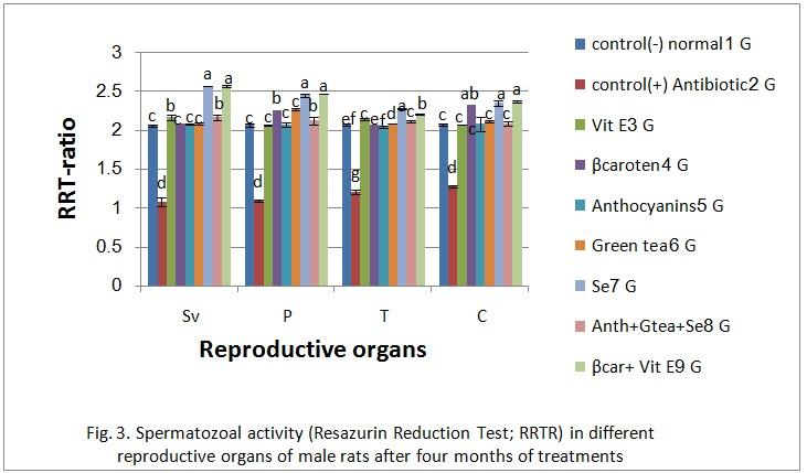
induces oxidative damage to rat testes, the antioxidant
Therefore, thespermatozoal activity was selected using
levels, testicular marker enzymes and marker of oxidative
resazurin reduction test (RRT), as a ratio between A580 to
stress were evaluated. The activity of SOD and CAT in the
A615 (RRTR). All groups, insignificant (p>0.05) alterations
post-mitochondrial fraction of rat testes decreased
were recorded in spermatozoal activity after treatment with
significantly by 31% and 53% in the rats treated with AZ
different antioxidants(Fig. 2). However, animals were
compared with the corresponding group of control rats (Fig
subjected to 20 mg AZ/kgbw showed significant (p<0.05)
4, 5). This decrease was prevented by 31% and 80%,
reduction in their spermatozoal activity. All treatments led to
respectively, on co-administration with Vit E. Treatment
significant (p<0.05) elevation in spermatozoal activity of all
with β-carotene increased the activity of SOD by 33% and
tested reproductive organs, except the treatment with
that of CAT by 92%. However co-administration with
selenium (G7) that led to insignificant change in
selenium (G7) or vitamin E + β-carotene (G9) recorded the
spermatozoal activity of caudaepididymes and testes (Fig.2).
highest elevation percent in the activity of SOD by 48%,
In general, the obtained data indicated that, the treatments
45% and that of CAT by 134% and 121%, but treatment
with selenium (G7) or vitamin E + β-carotene (G9)recorded
with anthocyanin (G5) and green tea (G6) recorded the
the highest value except only the treatment with selenium
lowest elevation percent in the activity of SOD by 25%for
(G7) that led to insignificant change in spermatozoal activity
each and that of CAT by 42% and 29%.
of testes, however treatment with selenium (G7), β-carotene
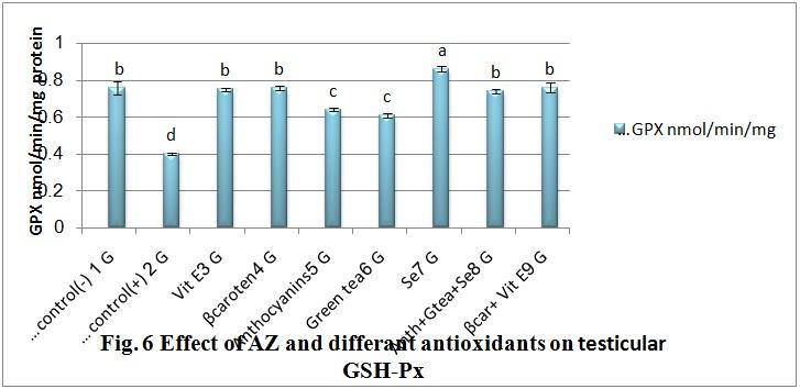
Testicular GSH-Px decreased significantly by 47%
(4) and it's mixture with vitamin E (G9) recorded the highest
in the AZ-treated rats relative to the controls (Fig. 6).All
value in caudaepididymes and (Fig.2). Moreover, the co-
treatment with antioxidants increased the level of the GSH-
admiration with different antioxidants led to attitude the
Px to normal except co-administration with selenium (G7)
hazard effect of Azithromycin on the spermatozoal activity.
that recorded the highest value in GSH-Px. In Contrary,
anthocyanin and green tea extracttreatments gave the lowest
elevation percent in the activity of GSH-Px.
Volume 2 2015 Issue 1 DEC-JAN AIJCSR
ISSN 2349 – 4425
www.americanij.com
revealed atrophy and degeneration of seminiferous tubules
associated with interstitial edema (Fig.8).
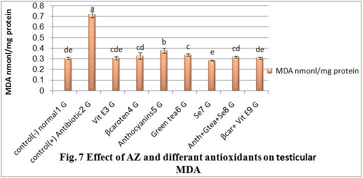
3.3.Markers of oxidative damage
The level of MDA, a marker of lipid peroxidation,
testicular MDA increased significantly (p<0.05) in rats
treated with AZ. These increment was markedly (p<0.05)
decreased by administration of all treatmentscomparing to
AZ-treated rats (Fig.7). Rationally, co-administration with
selenium (G7) recorded the lowest value in MDA.
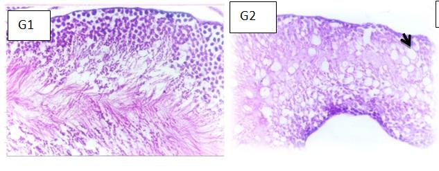
3.4. Histological changes
The photomicrographs in Fig. 8 illustrated the different
histopathologic changes that were observed in the testis of
animals given various treatments used in this work.
Administration of AZ caused severehistopathologic lesion
such as vacuolations and degeneration of spermatogoneal
cells lining seminiferous tubules. Normal seminiferous
tubules were noticed in testes of control negative rat (G1).
No histopathological changes were noticed in testes of rat
from group 3 (Fig.8). Increase diameter of seminiferous
tubule was noticed in testes of rat from groups 4 and 7
(Fig.8). However, examined sections from groups 5 and 6
revealed atrophy of seminiferous tubules (Fig.8). Apparent
normal seminiferous tubules were observed in testes of rats
from group 8 (Fig.8). Meanwhile, tests of rat from group 9
Volume 2 2015 Issue 1 DEC-JAN AIJCSR
ISSN 2349 – 4425
www.americanij.com
Fig (8) Histopathological image of Testes tissue from
control rats, AZ and AZ plus different antioxidants-treated
animals after in four months of treatments
G1:Control (-) (Normal control, basal diet), G2:Control
(+) (Control treated with 20mg kg-1bw of azithromycin),
G3:Vitamin E(3 IU /kg bw), G4:β-carotene (50mg/kg bw),
G5:Anthocyanin (71mg /kg bw),G6:Green tea extracted
(120mg/kg
bw),G8:[Anthocyanin (24mg /kg bw)+ , green tea extract
(40mg/kg bw) + Se (0.03mg/kg bw)], andG9: [Vitamin E(
1.5 IU /kg bw), +β-carotene (25mg/kg bw)].
Normal histopathological image of epididymis were
observed in all groups, except epididymis of control
negative rat (AZ administration) in group 2 showed
vacuolation of epithelial lining tubules (Fig.9).
Fig.9Histopathological image of epididymis tissue from
control rats , AZ and AZ plus different antioxidants-treated
animal after in four months of treatments
G1:Control (-) (Normal control, basal diet), G2:Control
(+) (Control treated with 20mg kg-1bw of azithromycin),
G3:Vitamin E(3 IU /kg bw), G4:β-carotene (50mg/kg bw),
G5:Anthocyanin (71mg /kg bw),G6:Green tea extracted
(120mg/kg
bw),G8:[Anthocyanin (24mg /kg bw)+ , green tea extract
(40mg/kg bw) + Se (0.03mg/kg bw)], andG9: [Vitamin E(
1.5 IU /kg bw), +β-carotene (25mg/kg bw)].
Volume 2 2015 Issue 1 DEC-JAN AIJCSR
ISSN 2349 – 4425
www.americanij.com
Microscopically, no histopatholgical change were noticed in
seminal vesicle of rat from group 1(control-ve) and group 4
(Fig.10). However, seminal vesicle of group 2 (control+ ve
rat) revealed atrophy and shortening of epithelial lining
(Fig.10). Meanwhile, seminal vesicle of rat from groups 3,
5, 7, and 9 showed hyperplasia of epithelial lining (Fig.10).
Examined sections of rat from group 6 showed focal
necrosis of epithelial lining (Fig.10). Marked atrophy and
necrosis of epithelial lining were observed in seminal vesicle
of rat group 8 (Fig.10).
Fig 10 Histopathological image of seminal vesicle
tissue from control rats, AZ and AZ plus different
antioxidants-treated animals after in four months of
G1:Control (-) (Normal control, basal diet), G2:Control (+) (Control
treated with 20mg kg-1bw of azithromycin), G3:Vitamin E(3 IU /kg bw),
G4:β-carotene (50mg/kg bw), G5:Anthocyanin (71mg /kg
bw),G6:Green tea extracted (120mg/kg bw),G7:Selenium (0.1 mg/kg
bw),G8:[Anthocyanin (24mg /kg bw)+ , green tea extract (40mg/kg bw) + Se
(0.03mg/kg bw)], andG9: [Vitamin E( 1.5 IU /kg bw), +β-carotene (25mg/kg
Volume 2 2015 Issue 1 DEC-JAN AIJCSR
ISSN 2349 – 4425
www.americanij.com
The microscopic investigation of prostate glandof group 2
(control+ ve rat) revealed necrosis associated with massive
leucocyte cells infiltration (Fig.11). Meanwhile prostate of
rat from group 1 (control-ve) and group 3 showed no
histopathological changes (Fig.11). However, prostate gland
of ratsfrom groups 4, 5 and 8 revealed hyperplasia and
vaculation of epithelial lining prostatic acini(Fig.11).
Meanwhile, examined sections from groups 6 and 7 showed
interstitial edema (Fig.11). On the other hand, prostate gland
of rat from group 9 revealed distension of the acini with
eosinophilic protein secretion(Fig.11) and hyperplasia of
epithelial lining some acini in another examined sections
from the same group 9(Fig.11).
Fig. 11 Histopathological image of Prostate tissue
from control rats, AZ and AZ plus different
antioxidants-treated animals after in four months of
treatments
G1:Control (-) (Normal control, basal diet), G2:Control (+) (Control
treated with 20mg kg-1bw of azithromycin), G3:Vitamin E(3 IU /kg bw),
G4:β-carotene (50mg/kg bw), G5:Anthocyanin (71mg /kg
bw),G6:Green tea extracted (120mg/kg bw),G7:Selenium (0.1 mg/kg
bw),G8:[Anthocyanin (24mg /kg bw)+ , green tea extract (40mg/kg bw) + Se
(0.03mg/kg bw)], andG9: *Vitamin E( 1.5 IU /kg bw), +β-carotene (25mg/kg
4. Discussion
A review of literature suggest that antimicrobial
agents can affect male reproductive functions including
sperm counts, motility, morphology, spermatogenesis and
germ cell integrity (35,29,52-55).Azithromycin is an azalide,
a subclass of macrolide antibiotics, structurally modified
from erythromycin. Its efficacy and potential to inhibit
microorganisms had led to its increased use. It is well-
known that Azithromycin is widely used to treat or prevent
certain bacterial infections, most often those causing middle
ear infections, strep throat, pneumonia, typhoid, and
Volume 2 2015 Issue 1 DEC-JAN AIJCSR
ISSN 2349 – 4425
www.americanij.com
sinusitis. AZ has been shown to induce a number of
serum testosterone (Table 2)may be AZ was able to reduce
biochemical disorders and increasing number of evidence
the rate limiting enzymes of testosterone synthesis,
indicates that it has risk of hepatotoxicity and cardio-toxicity
indicating a hindered production of the latter this isin
as adverse effect (34).This study appears to be the first
agreement with that of Ghosh&Dasupta (60) who reported
reporting the ability of the drug to cause oxidative damage
that gentamycin is aminoglycoside antibiotic make similar
to rats testicular function in rats.
For our Knowledge, this is the first time to reveal the AZ-
Regarding low fructose level in reproductive organs
induced oxidative stress, decreased antioxidant levels, lipid
(Fig. 2)seems to beconnected with the decreases in
peroxidation and histopathological changes in the testis,
testosterone levels. This explanation was also supposed by
epididymis and seminal vesicle. In the present study,
Melis (43),as fructose is the nutrient substance present in
administration of AZ did not affect the body weight of the
seminal plasma, and is initiated and controlled by testicular
animals but specifically caused a significant reduction in the
androgens (50). In this respect, hormonal deficiency causes
weights of testis, epididymis and seminal vesicle. The
a decrease or even disappearance of seminal fructose.In fact,
weight of the testis is largely dependent on the mass of the
treatment with androgens restores the ability of the
differentiated spermatogenic cells; the reduction in the
accessory glands to produce this sugar (61). Moreover,
weight of the testis may be due to decreased number of germ
fructose, when converted by spermatozoa to lactic acid
cells, inhibition of spermatogenesis and steroidogenic
provides an important source of energy for sperm cells. Our
enzyme activity (56).The reduction in testis and epididymis
studydemonstrated the relationship between the reduction in
weights is due to the marked parenchymal atrophy in
spermatozoal activity and the low level of fructose (Fig. 2
germinal cell layer thickness and the other deteriorated
and Fig. 3)as statedby earlier studies (62)as well as other
histopathologic findings in testis, along with decreased RRT
researchers Melis (43). The only parameter that appears to
ratio, which were observed in AZ-treated rats in the present
be possibly improved with oral antioxidant therapy is sperm
study. Similar findings were reported earlier by Turk et al.,
motility. Many studies have shown small but significant
(57).The observed weight loss of the accessory sex organs
improvements in sperm motility with supplementation of
may be due to reduced bioavailability of sex hormones such
selenium (63), Vitamin E (64), Vitamin E and selenium
as testosterone, luteinizing hormone, follicle stimulating
(65), glutathione (66) and Astaxanthin(67), as well
hormone and inhibit B, the levels of which in the circulation
Many years ago,MacLeod (68) noted that
indicate the reproductive endocrine status of the male as has
incubation of sperm under conditions of high oxygen tension
been stated beforewith Schrade, (58). All antioxidants
lead to a rapid loss of their motility. The addition of the
administrated significantly mitigated the effect of AZ on the
antioxidant catalase to the medium preserved sperm motility
testicular weight and other accessory reproductive organs
prompting MacLeod (68)to suggest that sperm must
affected by the drug.More recently Elias & Nelson, (55)
produce hydrogen peroxide during normal oxidative
have emphasized that testosterone is very essential in
metabolism. Since this publication, it has evolved that three
spermatogenesis. It was reported that testosterone is
inter-related mechanisms account for oxidative stress-
essential in growth and association with follicle stimulating
mediated male infertility—impaired motility, impaired
hormone acts on the somniferous tubules to initiate and
fertilization and oxidative DNA damage.
maintain spermatogenesis. Earlier, Louvetet al., (59)point
In normal situation the seminal plasma contains antioxidant
out that interstitial cell-stimulatinghormone (ICSH), also
mechanisms which are likely to quench these ROS and
known as Lutenizing hormone (LH) stimulates Leydig cell
protect against the damage to spermatozoa(29). It well
production of testosterone. In accordingly, the level of these
known that bactericidal antibiotics can induce cellular death
two sexual hormones; testosterone (T) and ICSH were
through a common oxidative damage mechanism that relies
determined in blood serum of the investigated rats (Table
on the production of ROS. Through their various primary
2).Azithromycin administration significantly decreased
targets, antibiotics can activate cellular respiration, which
Volume 2 2015 Issue 1 DEC-JAN AIJCSR
ISSN 2349 – 4425
www.americanij.com
leads to the formation of superoxide and the release of iron
mg of Vitamin E and 225 mg of selenium (65) or 300 mg of
from iron-sulfur clusters. Free iron then activates a chemical
Vitamin E alone (64) have been shown in placebo controlled
reaction (the Fenton reaction) to produce ROS in the form of
studies to reduce sperm MDA levels. Finally, a well-
hydroxyl radicals (OH•) (30).The administration of AZ
designed RCT of 2 months treatment with 1 g of Vitamin C
induced marked oxidative stress and altered the antioxidant
and Vitamin E reported a very significant reduction in sperm
status in rats and its mechanism of toxicity appears to
DNA damage (75). This finding is supported by non-
precede through the generation of free radicals or depletion
controlled studies also reported a reduction in sperm DNA
of the antioxidant systems(34).
damage with the use of a combination of Vitamin C and E
In connection, animals treated with AZ in this study
(400 mg each), β-carotene (18 mg), zinc and selenium (76)
showed decreased activities of antioxidant enzymes
otherwise a combination of acetylcysteine, 180 mg Vitamin
SOD,CAT and GSH-PX in the testis. This decrease in the
E, 30 mg β-carotene and essential fatty acids (73).
antioxidant defense molecules led to a concomitant increase
Various agents have been attempted to protect from
in the level of MDA. The inactivation of the antioxidant
and/or prevent the side effects of many chemotherapeutics.
enzymes may be caused by excess ROS generated in the
Flavonoids are among these agents and are found in almost
system (69). Our data concluded that co-administration of
all food categories such as fruits and vegetables being the
Vit E, β-carotene, anthocyanin, green tea extract, mixture of
main source. Flavonoids have many functions, such as
the (anthocyanins+ green tea extract+ Se) or mixture of the
phenolic antioxidants, scavengers of free radicals, chelating
(Vitamin E+ β-carotene) with AZ significantly attenuated
agents, and modifiers of various enzymatic and biologic
the effects of AZ drug on the antioxidant enzymes and
reactions (77). Anthocyanins and green teais a naturally
further suggests that impairment of male fertility through
occurring plant polyphenol that exhibits antioxidative
induction of oxidative stress. Olayinka&Ore (34)observed
properties both in vivo and in vitro. In fact, they have been
a significant reduction in the activities of SOD, CAT and
shown to exert a potent scavenging action on both O -
GST in the liver of azithromycin- treated animals. The
˙OH, as well as lipid peroxidationbeside increase the
significantly decreased activity of the ROS scavenging
activities of SOD and Catalase(78- 80).
enzyme, SOD and CAT, by exposure to azithromycin,
The declining of lipid peroxidation in all samples
conforms to previous report on macrolide antibiotics (70).
studied apparently indicates that all treatments potently
This may be due to the damaging effects of free radicals
scavenged the free radicals (O2 - and ˙OH), and suppressed
possibly generated by the action of the drug a clear oxidative
oxidative DNA damage. The antioxidant activity of Vit E, β-
stress has been achieved in the testis. Lipid peroxidation is a
carotene, anthocyanin, green tea,
process of oxidative degredation of polyunsaturated fatty
(anthocyanin+ green tea extract + selenium) or mixture of
acids that result in impaired membrane structure and
the (Vitamin E+ β-carotene) and mitigation of ROS-
function (71).
induced depletion of GSH-Px and CAT activities also show
In the main line this prospectus, several studies
that all used antioxidants in the present study especially Se,
have reported that levels of ROS within semen can be
Vitamin E and β-carotenehas strong antioxidant activity.
reduced by augmenting the scavenging capacity of seminal
The proposed mechanism of action within which
plasma using oral antioxidant supplements. The oral
may profoundly includethe non-enzymatic antioxidants
antioxidant Astaxanthin(67), carnitine(72) or a combination
present within semen include -tocopherol (Vitamin E),
of antioxidants such as acetylcysteine, β-carotene, Vitamin
glutathione, carotenoids, and flavonoids. These agents
E and essential fatty acids (73) directlybinding cure with
principally act by directly neutralizing free radical activity
reduce seminal ROS levels. likewise,a randomized control
chemically. However, they also provide protection against
study comparing 3 months of Vitamin E (600 mg/day)
free radical attack by two other mechanisms. Albumin in
treatment with placebo has confirmed this reduction in
another nutrient system can intercept free radicals by
seminal ROS levels (74). Furthermore, a combination of 400
Volume 2 2015 Issue 1 DEC-JAN AIJCSR
ISSN 2349 – 4425
www.americanij.com
becoming oxidized itself, thereby sparing sperm from
Free radicals, otherwise damage cellular lipids by
attack(81).
binding to membrane anionic phospholipids (34), proteins
Dietary deficiencies have been linked with sperm
and DNA, finally the entire cell (88). Antioxidants, being
oxidative damage as stated by several research works. The
free radical scavengers hamper the destructive effects of the
Age and Genetic Effects in Sperm (AGES) study examined
latter on cells and tissues. AZ-induced renal and hepatic
the self-reported dietary intake of various antioxidants and
toxicity in rats as explained by the photomicrograph of rats
nutrients (vitamins C and E, β-carotene, folate and zinc) in a
kidney and liver sections. The histological alterations
group of 97 healthy nonsmokers and correlated this with
observed in the liver of azithromycin - treated rats
sperm quality (82). The pre study reported a significant
characterized by sinusoidal and portal congestion, and mild
correlation between vitamin C intake and sperm
periportal cellular infiltration by mononuclear cells coupled
concentration and between vitamin E intake and total
with the presence of protein casts in the kidney tubular
progressively motile sperm. This is also consistent with
lumen as well as severe cortical congestion and hemorrhage
earlier reports of a significant link between seminal plasma
is an indication of disruption of cellular architecture. All
vitamin E levels and an increase in percentage of motile
these abnormalities might have resulted due to the formation
sperm (83).Alternatively, differences in the populations
of highly reactive radicals caused by the drug which
studied may explain the discrepant results. Song et al., (84)
disrupted normal cellular functioning of the liver and the
correlated sperm DNA damage with dietary antioxidant
kidney(34). This phenomenon would have been responsible
intake in infertile men, while Silver et al., (85)examined this
for structural changes such as vacuoles, gaps, cytotoxicity
relationship in healthy presumed fertile patients. Fertile men
and atrophic changes in the testis and the other parts of the
with low levels of oxidative attack may not be as dependent
reproductive system (accessory sex organs). The vacuoles
on seminal antioxidants for protection of their sperm DNA
and gaps in the seminiferous epithelium appear due to
integrity. Therefore, a dietary deficiency in antioxidants may
dilatation of endoplasmic reticulum in Sertoli cells (89), and
not lead to sperm oxidative DNA damage in this fertile
missing of germ cells from the epithelium (90), respectively.
The oxidatative stress in the testis should have affected the
While many studies have shown improvements in sperm
structure and functions of cell organelles that manifested as
quality with antioxidant treatment, the ability of these
histopathological changes. Since the oxidative stress-status
changes to translate into improved chances of pregnancy is
induces cellular and DNA damage (91), it is possible that
less clear. The Menevitnutraceutical is postulated to improve
the cell death and consequent tubular atrophy, at least in a
sperm quality by three complimentary mechanisms. First, it
few tubules, would have been the response of seminiferous
contains traditional antioxidants such as Vitamins C and E,
epithelium to altered biochemical milieu in the testis. These
selenium and lycopene to protect sperm from ROS already
results therefore indicate that the oxidative stress induced
produced. Second, it contains garlic which is known to have
owing to an imbalance between the free radicals and
an anti-inflammatory effect, thereby potentially reducing
oxidants, has a key role in initiating tubular damage in the
seminal leukocyte ROS production (86). Finally, Menevit
testis, just as it does in the liver and kidneys (34).
contains zinc, selenium and folate that are believed to play a
Taken together, the present study reveals
role in augmenting protamine packaging of sperm DNA
thatadministration of therapeutic dose of AZ tomale rats
(87), helping to protect sperm from ROS attack. While it is
induced oxidative stress by decreasing the antioxidant
yet to be proven that combinational therapy such as
system, increased lipid peroxidation and concomitantly
Menevitimproves sperm DNA integrity, it appears logical
impairs testicular function, spermatogenesis and epididymal
that using several antioxidants with different modes of
sperm physiology and morphology. All antioxidants
action, together with an agent to reduce leukocyte ROS
administrated in this study exhibited similarities in their
production (23) is most likely to result in a beneficial effect.
capability to mitigate the toxic responses of AZ, which
suggests that the adverse effects of AZ on the testes are at
Volume 2 2015 Issue 1 DEC-JAN AIJCSR
ISSN 2349 – 4425
www.americanij.com
least in part due to impairment of the antioxidant defense
[ 3 ] Martini M.C. Infertility. In: PernollM,l.
system and further enhancement of lipid peroxidation. The
ed. Current obstetric and gynecologic diagnosis
inability of these antioxidants to fully protect the testes
and treatment. Reproductive endocrinology and
against AZ-induced toxicity suggests that the antibiotic
infertility.7th ed. Norwalk (CT): Appleton &
could mediate testicular damage through other mechanisms
apart from oxidative stress. In view of the importance of this
Lange.1991; 1025-1036.
drug in clinical practice, the relevance of our study to
[ 4 ] Sigman M, Lipshultz L.I., Howards
humans merits further investigation on the mechanisms by
which AZ causes testicular toxicity.
S.S.Evaluation of the subfertile male.In: Lipshultz
In Conclusion, The long term use of azithromycin
LI, Howards SS, eds. Infertility in the male. St.
in the treatment of bacterial prostatitis produced good
Louis: Mosby-Year Book, Inc.1997; 173-93.
outcome but limited information on toxicological effect on
testicular function could limited it long term use. This study
[ 5 ] Remah M KamelManagement of the
apparently suggests that all used antioxidants have a
infertile couple: an evidence-based protocol.
protective effect against testicular toxicity caused by AZ.
Reproductive Biology and Endocrinology, 2010;
Therefore, co-administrated antioxidants may be used
8:21 doi:10.1186/1477-7827-8-21
combined with AZ in chemotherapeutic treatments to
improve AZ-induced injuries in sperm quality and oxidative
[ 6 ] Swan SH Does our environment affect our
stress parameters.
fertility? Some examples to help reframe the
question. SeminReprod Med, 2006;24:142-146.
I would like to thank Prof. DR. Ashraf Abd-
Elwahed , Professor of pediatrics, Faculty of medicine Ain
[ 7 ] Kolettis PN Evaluation of the sub fertile
Shams University, For giving every possible advice
man Am, Fam. Physician, 2003; 68: 1265- 2172.
throughout the investigation and during writing of the
[ 8 ] Schlegel
I want also to thank Prof. Dr. KawkabAbd El-Aziz
Marshall, F.F. Antibiotics: potential hazards to
Ahmed, Professor of Pathology Department, Faculty of
Veterinary Medicine, Cairo University, for her valuable
male fertility. Fertil. Steril.,1991; 55, 235–242.
discussions and explain the obtained results.
[ 9 ] Onyije F.M. Drug: A Major Cause of
Infertility in Male Asian J. Med. Pharm.
Res.,2012; 2(2): 30-37.
[ 1 ] Nieschlag E. Scope and goals of
Andrology. In: Andrology - male reproductive
[ 10 ] Hargreaves CA., S Rogers, F Hills, F
health and dysfunction. Nieschlag E and Behre
Rahman, JS Howell Richard and ST Homa.
HM. (eds). Springer Verlag, Berlin, Federal
co-trimoxazole,
erythromycin,
Republic of Germany.1997; pp. 1-8.
amoxycillin, tetracycline and chloroquine on
sperm function in vitroHuman Reproduction
[ 2 ] World
Statistics
1998; 13:1878–1886.
(2013)Population
Division,
Department
Economic and Social Affairs, United Nations,
[ 11 ] Sharma R, Biedenharn KR, M JF
World Population Prospects: The 2012 Revision
&Agarwal A Lifestyle factors and reproductive
Volume 2 2015 Issue 1 DEC-JAN AIJCSR
ISSN 2349 – 4425
www.americanij.com
fertility
[ 18 ] Retsema JA, Girard W, Schelkly M,
Reproductive
Manousos M, Anderson G, Bright R, Mason R.
Endocrinology2013;11:66, 1-15.
Spectrum and mode of action of azithromycin
(CP-62,993), a new 15 membered-ring macrolide
[ 12 ] Andrews,
Bavister,
with improved potency against gramnegative
Capacitation of hamster spermatozoa with the
organisms.
divalent cationchelators D-penicillamine, L-
histidine,and L-cysteine in a protein free culture
medium. Gamete Res., 1989; 23, 159–170.
[ 19 ] Er A, Ulutas E, Altan F, Cetin G, Bulbul A,
Elmas M, Yazar E. Tulathromycin disturbs blood
[ 13 ] Adikwu E. and Brambaifa N. Toxicological
oxidative and coagulation status. Afr. J.
Effect of Perfloxacin on Testicular Function of
Biotechnol.2011; 10(16):3243-3247.
Male Guinea Pigs. Asian j. Exp. Biol. Sci.
2012;Vol 3 (1): 28 – 33.
[ 20 ] Lockwood AM, Cole S, Rabinovich M.
Azithromycin-induced liver injury. Am J Health
[ 14 ] Jerry M, and Zuckerman MD. Macrolides
Syst Pharm. 2010; 67(10):810-814.
ketolides:
telithromycin. Infect Dis Clin N Am. 2004;
[ 21 ] Kumar V, Harjai K, and Chhibber S. Effect
18:621–649.
of clarithromycin on lung inflammation and
macrpghage
[ 15 ] Piscitelli SC, Danziger LH, and Rodvold
Klepsiellapenumoniae B5055-induced acute lung
KA. Clarithromycin and azithromycin: new
infection in Balb/C mice.J Chemother. 2008;
macrolide antibiotics. Clin Pharm.1992;11:137–
20:609-614.
[ 22 ] Gupta S, Agarwal A, Banerjee J, and
[ 16 ] Zuckerman JM, Qamar F, and Bono BR
Alvarez JG. The role of oxidative stress in
Macrolides,
ketolides,
glycylcyclines:
spontaneous abortion and recurrent pregnancy
azithromycin,
clarithromycin,
telithromycin,
tigecycline.Infectious Disease Clinics of North
[ 23 ] Tremellen K Oxidative stress and male
[ 17 ] Klausner
Jeffrey D,
PassaroDRJ,
infertility—a
perspective
Thacker WL, Talkington DF, Werner SB, and
Reproduction Update,2008; 14: 243–258.
Vugia, Duc J. Enhanced Control of an Outbreak
of Mycoplasma pneumonia with Azithromycin
[ 24 ] Oliva R. Protamines and male infertility.
Prophylaxis.The
Infectious
Hum Reprod Update2006;12:417– 435. 25.
Diseases 1998; 177 (1): 161–166.
[ 25 ] Villegas J, Schulz M, Soto L, Iglesias T,
Miska W, Sanchez R. Influence of reactive oxygen
Volume 2 2015 Issue 1 DEC-JAN AIJCSR
ISSN 2349 – 4425
www.americanij.com
species produced by activated leukocytes at the
[ 34 ] Olayinka ET and Ore A.Influence of
apoptosis
Azithromycin
Treatment
on Hepatic Lipid
spermatozoa.FertilSteril2005; 83: 808–810.
Peroxidation and Antioxidant Defence Systems of
RatsBritish Journal of Pharmaceutical Research
[ 26 ] Wolff H, Politech JA, Martinez A,
2014; 4(2): 240-256.
Haimovici F, Hill JA and Anderson AJ
Leukocytospermia is associated with poor semen
[ 35 ] Raji Y, Olufadekemi T, Salami SA, and
quality. Fertile and Sterile 1990; 53, 528-536.
Bolarinwa AF Impact of -tocopherol on
metronidazole and tetracycline-induced alteration
[ 27 ] Nicol, S. G. Pro-oxidative and antioxidant
in reproductive activities of male albino
imbalance in human semen and its relation with
rats.Journal of Biological Science 2007; 7(1):41-
male infertility. Asian J. Androl. 2004;6, 54-65.
[ 28 ] Yadav, S.B., Sardeshamukh, A.S. and
[ 36 ] Hu Q, Pan G, and Zhu J Effect of
Suryakar, A.N. A study of seminal hyaluronidase,
fertilization on selenium content of tea and the
fructose, lipid peroxide and Zinc in primary male
nutritional function of Se-enriched tea in rats
infertility. Journal of Obst. and Gyn. of India
Plant and Soil. 2002; 91: 91-95.
2001;51 (5),142-145.
[ 37 ] Revilla E, Ryan JM and Oretga GM
[ 29 ] Yadav SB, Suryakar AN, Huddedar AD
Comparison of several procedures used for the
and Shukla PS Effect of antioxidants and
extraction of Anthocyanins from red grapes J.
antibiotics on levels of seminal oxidative stress in
Agric. Food Chem. 1998; 46: 4592 - 4597.
leukocytespermic infertile men Indian Journal of
Clinical Biochemistry, 2006; 21 (1) 152-156.
[ 38 ] El-DakakAbeer M Effect of natural
antioxidants in grapes and carrots on cholesterol
[ 30 ] Belenky P & Collins J Antioxidant
levels Thesis of Ph.D.Agric. 2004; (Food Sci&
strategies to tolerate Antibiotics Science 2011; vol
Tech.), Cairo University.
334, 18 NOVEMBER pp.915.
[ 39 ] Chen BH and Tang YC Processing and
[ 31 ] Nguyen D., Science 2011; 334, 982
stability of carotenoid powder from carrot pulpJ.
[ 32 ] Goswami M., Mangoli S.H. &Jawali N.
Agric. Food Chem. 1998; 46: 2312 - 2318.
Antibiotics and antioxidants: Friends or Foes
[ 40 ] El-DakakaAbeer M, Ahmed Mona H, and
during Therapy? Feature Article, ISSUE NO.
Ismail SA Effect of some natural antioxidants on
2011;323 I NOV. – DEC, 42-46.
oxysterol
endogenously
[ 33 ] Cerny A Side effects and consequences of
frequently
antibiotics
Biomedical Sciences2008;28: 85- 102.
ft. J. Suisse de Medizinische,1996; 126: 528- 534.
Volume 2 2015 Issue 1 DEC-JAN AIJCSR
ISSN 2349 – 4425
www.americanij.com
[ 41 ] Zamora R, Hidalgo F and Tappel L
selenium influences the response to fatty acid-
Comparative antioxidant effectiveness of dietary
induced oxidative-stress in humans.Biological
β-carotene, vitamin E, selenium and Coenzyme
Trace Element Research 1997; 60: 51-60.
Q10 in rat erythrocytes and plasmaJ. Nutr.1991;
[ 50 ] Mann T.Fructose and fructolysis in semen
121: 50-56.
in relation to fertilityLancet i, 1948; 446–448.
[ 42 ] Bajaj VK and Gupta RS Fertility
[ 51 ] Bancroft, J.D., Stevans, A., D.R., T.Theory
suppression in male albino rats by administration
and practice of histological techniques, 4 ed.
methanolic
Opuntiadillenii.
Churchill
Livinigstone,
Edinburgh,
Andrologia2012;44, 530–537.
Melbourne, New York., 1996.
[ 43 ] Melis MS Effects of chronic administration
[ 52 ] Olayemi FO Review: A review on some
of Stevia rebaudiana on fertility in rats Journal of
causes of male infertility African Journal of
Ethnopharmacology1999; 167:157–161.
Biotechnology 2010;9(20) 2834-2842.
[ 44 ] Reddy,
Bordekar,
[ 53 ] Farombi
Ugwuezunmbaa
resazurin
Ezenwadua TT., Oyeyemib MO, and Ekor
reduction test and semen quality in men. Indian J.
M.Tetracycline-induced reproductive toxicity in
Exp. Biol.1999; 37, 782-786.
male rats: Effects of vitamin C and N-
[ 45 ] Gornall AC and Bardawill CJ J. Biol.
Toxicologic
Chem.1949; 177: 751.
Pathology2008; 60: 77–85.
[ 46 ] Nishikimi, M.; N.A., Roa and K.,YogThe
[ 54 ] Zobeiri F, Sadrkhanlou RA, Salami S,
occurrence of superoxide anion in the reaction of
Mardani K, Ahmadi A.The effect of ciprofloxacin
reduced phenazinemethosulphate and molecular
on sperm DNA damage, fertility potential and
oxygen.Biochem. Bioph. Res. Common.1972; 46:
early embryonic development in NMRI mice
849 -854.
Veterinary Research Forum. 2012; 3 (2) 131 –
[ 47 ] Aebi, H.Catalase in vitro. Methods
Enzymol.1984; 105 : 121 – 136.
[ 55 ] Elias A. and Nelson B. Toxicological effect
of Ciprofloxacin on testicular function of male
[ 48 ] Waschulewski, I.H. and Sunde, R.A. Effect
guinea pigs Asian J. Exp. Biol. 2012; Vol.
of dietary methionine on utilization of tissue
3(2):384-390.
selenium from dietary selenomethionine for
glutathione peroxidase in the rat.J. Nutr. 1988;
[ 56 ] Takahashi O and Oishi S. Testicular
118: 367-374.
toxicity of dietary 2, 2-bis (4-hydroxyphenyl)
propane (bisphenol A) in F344 rat.Arch Toxicol
[ 49 ] Metlzer, H.M., M., Folmer; S., Wang; Q.,
2001; 75:42–51.
Lie; A., Maggi and H.H., MundalSupplementary
Volume 2 2015 Issue 1 DEC-JAN AIJCSR
ISSN 2349 – 4425
www.americanij.com
[ 57 ] Turk G, Atessahin A, Sonmez M, Ceribasxi
[ 64 ] Suleiman SA, Ali ME, Zaki ZM, el-Malik
AO and YuceAImprovement of cisplatin-induced
EM, Nasr MA. Lipid peroxidation and human
injuries to sperm quality, the oxidant-antioxidant
sperm motility: protective role of vitamin E. J
system, and the histologic structure of the rat
Androl 1996; 17:530–537.
testis by ellagic acid. Fertility and Sterility
[ 65 ] Keskes-Ammar L, Feki-Chakroun N, Rebai
2008;89 (5):1474-1481.
T, Sahnoun Z, Ghozzi H, Hammami S, Zghal K,
[ 58 ] Schrade SM. Man and the workplace:
Fki H, Damak J, Bahloul A. Sperm oxidative
assessing his reproductive health.Chem Health
stress and the effect of an oral vitamin E and
Saf 2003; 11–16.
selenium supplement on semen quality in infertile
men. Arch Androl 2003; 49:83–94.
[ 59 ] Louvet J, Harman S, and Ross G Effects of
chorionic
gonadotropin,
[ 66 ] Lenzi A, Culasso F, Gandini L, Lombardo
ninterstitial cell stimulating hormone and human
F, Dondero F. Placebo-controlled, double-blind,
follicle-stimulating hormone on ovarian weights
cross-over trial of glutathione therapy in male
in estrogen-primed hypophysectomized immature
infertility.Hum Reprod 1993; 8:1657–1662.
female rats. Endocrinol.1975; 96 1179-1186.
[ 67 ] Comhaire FH, El Garem Y, Mahmoud A,
[ 60 ] Ghosh S and Dasupta S.Gentamicin
Schoonjans
induced inhibition of steroidogenic enzymes in rat
conventional/antioxidant ‘Astaxanthin' treatment
testis.Indian. J. Physiol. Pharmacol.1999; 43,
for male infertility: a double blind, randomized
247- 250.
trial. Asian J Androl 2005; 7: 257–262.
[ 61 ] Kempinas WG and Lamano-Carvalho
[ 68 ] MacLeod J. The role of oxygen in the
TL.A method for estimating the concentration of
metabolism and motility of human spermatozoa.
spermatozoa in the rat cauda epididymis Lab.
Am JPhysiol 1943; 138:512–518.
Anim.1988;22, 154-156.
[ 69 ] Pigeolet E, Corbisier P, Houbion A,
[ 62 ] El-DakakaAbeer M, KhirallaGhada M, El-
Lambert D, Michiels DC, and Raes M.Glutathione
Nahal Dalia M. Effect of partial replacement of
peroxidase, superoxide dismutase and catalase
monosodium
glutamate
5΄-inosine
inactivation by peroxides and oxygenderived free
monophosphate on the fertility of male rats Arab
radicals. Mech Ageing Dev 1990;51:283–290.
Universities J. of Agric. Sci 2014;Vol. 22 (2): 409-
[ 70 ] Yazar E, Altunok V, Elmas M, Tras B, Bas
AL, Ozdemir V. Effect of tilmicosin on cardiac
[ 63 ] Scott R, MacPherson A, Yates RW,
superoxide dismutase and peroxidase activities.J
Hussain B, Dixon J. The effect of oral selenium
Vet Med B.2002; 49: 209-210.
supplementation on human sperm motility.Br J
Urol 1998;82:76–80.
Volume 2 2015 Issue 1 DEC-JAN AIJCSR
ISSN 2349 – 4425
www.americanij.com
[ 71 ] Goel A, Dani V and Dawan DK.protective
quercetin in mice Food ChemToxicol.1999;37
effects of zinc on lipid peroxidation, antioxidant
:313–318.
[ 78 ] Jeong YJ, Choi YI, Kwon HM, Park HS,
chlorpyrifos induced toxicity. Chemico. Biol.
Lee M and Kang YH.Differential inhibition of
Inter. 2005; 156,131- 140.
LDL-indued
apoptosis
[ 72 ] VicariE, and Calogero AE. Effects of
endothelial
different
treatment with carnitines in infertile patients with
flavonoidsBritish Journal of Nutrition2005; 93:
581- 591.
[ 79 ] Yokozawa T, Nakagawa T, and Kitani
[ 73 ] Comhaire FH, Christophe AB, Zalata AA,
K.Antioxidative activity of green tea polyphenol in
Dhooge WS, Mahmoud AM, Depuydt CE. The
cholesterol-fed ratsJ. Agric. Food Chem.2003;
effects of combined conventional treatment, oral
50: 3549- 3552.
antioxidants and essential fatty acids on sperm
[ 80 ] Turk G, Atessahin A, Sonmez M, Aydin M,
subfertile
Prostaglandins
Yuce A, Gur S, Yuksel M, Aksu EH, and Aksoy
LeukotEssent Fatty Acids 2000; 63:159–165.
H.Effects of pomegranate juice consumption on
[ 74 ] Kessopoulou E, Powers HJ, Sharma KK,
sperm quality, spermatogenic cell density,
Pearson MJ, Russell JM, Cooke ID, Barratt CL. A
antioxidant activity and testosterone level in male
double-blind randomized placebo cross-over
rats. Clinical Nutrition 2008;27, 289-296.
controlled trial using the antioxidant vitamin E to
[ 81 ] Twigg J, Fulton N, Gomez E, Irvine DS,
treat reactive oxygen species associated male
Aitken RJ. Analysis of the impact of intracellular
infertility.FertilSteril 1995; 64:825–831.
reactive oxygen species generation on the
[ 75 ] Greco E, Iacobelli M, Rienzi L, Ubaldi F,
structural and functional integrity of human
Ferrero S, Tesarik J. Reduction of the incidence of
spermatozoa:
peroxidation,
sperm DNA fragmentation by oral antioxidant
fragmentation and effectiveness of antioxidants.
treatment. J Androl 2005; 26:349–353.
Hum Reprod1998;13:1429–1436.
[ 76 ] Menezo YJ, Hazout A, Panteix G, Robert
[ 82 ] Eskenazi B, Kidd SA, Marks AR, Sloter E,
F, Rollet J, Cohen-Bacrie P, Chapuis F, Clement
Block G, Wyrobek AJ. Antioxidant intake is
P, Benkhalifa M. Antioxidants to reduce sperm
associated with semen quality in healthy men.
DNA fragmentation: an unexpected adverse effect.
Hum Reprod 2005; 20:1006–1012.
Reprod Biomed Online2007; 14:418–421.
[ 83 ] Therond P, Auger J, Legrand A, Jouannet
[ 77 ] Khanduja KL, Gandhi RK, Pathania V,
P. alpha-Tocopherol in human spermatozoa and
and Syal N.Prevention of N-nitrosodiethylamine–
seminal plasma: relationships with motility,
induced lung tumorigenesis by ellagic acid and
Volume 2 2015 Issue 1 DEC-JAN AIJCSR
ISSN 2349 – 4425
www.americanij.com
antioxidant enzymes and leukocytes.Mol Hum
the male Wistar rat.Folia. Morphol 2005, 64, 65-
Reprod 1996; 2:739–744.
[ 84 ] Song
[ 91 ] Tramer F, Rocco F,Micali F, Sandri G and
Relationship between seminal ascorbic acid and
Panfili E.Antioxidant systems in rat epididymal
sperm DNA integrity in infertile men. Int J Androl
spermatozoa. Biol. Reprod.1998; 59, 753- 758.
2006; 29:569–575.
[ 85 ] Silver EW, Eskenazi B, Evenson DP, Block
G, Young S, Wyrobek AJ. Effect of antioxidant
intake on sperm chromatin stability in healthy
nonsmoking men. J Androl 2005; 26:550–556.
[ 86 ] Chang HP, Huang SY, Chen YH.
Modulation of cytokine secretion by garlic oil
derivatives is associated with suppressed nitric
oxide production in stimulated macrophages. J
Agric Food Chem 2005; 53:2530–2534.
[ 87 ] Pfeifer H, Conrad M, Roethlein D,
Kyriakopoulos A, Brielmeier M, Bornkamm GW,
Behne D. Identification of a specific sperm nuclei
selenoenzyme necessary for protamine thiol cross-
linking during sperm maturation.FASEB J. 2001;
15:1236–1238.
[ 88 ] Mates JM, Perez-Gomez C and Nunez
DCI.Antioxidant enzymes and human disease.
Clin. Biochem. 1999; 32, 595- 603.
[ 89 ] De Krester DM and Kerr JB. The cytology
of the testis. In (Knobil NA and Neil JD., eds.),
pp.1177- 1280. The physiology of reproduction.
2nd Ed. Raven Press Ltd. New York 1994.
[ 90 ] Narayana K, D'Souza UJA, Prashanthi N
and Ganesh K. The antiviral drug ribavirin
reversibly affects the reproductive parameters in
Volume 2 2015 Issue 1 DEC-JAN AIJCSR
Source: http://yloop.in/index.php/AIJ/article/download/54/54
History of Buddhism HISTORY OF BUDDHISM SRI In order to understand and appreciate the history of early Buddhism in Sri Lanka we should have, as a background some general idea of the India of the third century B.C from where Buddhism come to Sri Lanka and also of the pre-Buddhist Sri Lanka to which it was introduced. When the Indian missionaries brought Buddhism to this Island they carried here with them not only the teaching of the Buddha but also the culture and civilisation of Buddhist India. Almost all Buddhist rites , ceremonies, festivals and observance of Sri Lanka were with slight local changes and modification, the continuation of Indian practice which the early Buddhist missionaries introduced into this country. It necessary there at the time of the advent of Buddhism to Sri Lanka.
CONTRATO RED HAT ENTERPRISE AGREEMENT This Red Hat Enterprise Agreement, including all referenced El presente Contrato Red Hat para Empresas, incluidos todos los anexos appendices and documents located at URLs (the "Agreement"), is a que se ha hecho referencia y documentos ubicados en los URL (en between Red Hat Limited ("Red Hat") and the purchaser or user of