Effectiveness of intralesional triamcinolone in the treatment of keloids in children

Pediatric Dermatology Vol. 33 No. 1 75–79, 2016 Effectiveness of Intralesional Triamcinolone in the Treatment of Keloids in Children Silvana Acosta, M.D.,* Ester Ureta, M.D.,† Ricardo Ya nez, M.D.,* Natalia Oliva, M.D.,‡ Susana Searle, M.D.,* and Claudio Guerra, M.D.* *Plastic Surgery Section, Surgery Division, Medical School, Pontificia Universidad Cat olica de Chile, Santiago, Chile, †Radiology Unit, Clınica Alemana, Puerto Varas, Chile, ‡Faculty of Chemistry, School of Pharmacy, Pontificia Universidad Cat olica de Chile, Santiago, Chile Background: The current treatment of keloids includes surgery, intralesionalsteroids, and radiotherapy, among others. Radiotherapy is not recommended inchildren due to its effects on growing tissues. Our aim was to study intralesionaltriamcinilone therapy of keloids in children and analyze the impact of bodylocation, age of the lesion, and etiology of the keloid on clinical response.
Methods: We conducted a prospective clinical trial with patients 1 to 14 years ofage evaluated for keloid treatment. A soft tissue ultrasound was performed tomeasure the keloid volume, prior to intralesional infiltration with triamcinoloneacetonide. A posttreatment ultrasound quantified the volume differencesattributed to therapy. For the analysis, Mann–Whitney/Wilcoxon test for pairedsamples and a multiple regression analysis were performed.
Results: Twenty-one patients with a total of 25 keloids were enrolled, with amedian age of 12 years (range 6–14 yrs). The initial lesional volume was 1.25 cc(range 0.2–6.3 cc) and the final volume was 0.2 cc (range 0.0–1.53 cc),corresponding to 82.7% of size reduction (p < 0.001). Regarding the rela-tionships between response and body location, etiology and age of the lesion,the multiple regression analyses obtained p-values of 0.46, 0.16, and 0.87,respectively. One patient failed to improve. Average follow-up was 30 months.
Conclusions: Triamcinolone acetonide is highly effective for the treatment ofpediatric keloids. There is no relationship between clinical response and thefactors evaluated, such as lesion location, etiology and age of the keloid.
Address correspondence to Silvana Acosta M.D.,, Secci Cirugıa Plastica, Divisi on de Cirugıa, Escuela de Medicina, Pon- tificia Universidad Cat olica de Chile, Marcoleta 350, Santiago 8330024, Chile, or e-mail: [email protected].
This study was presented at "LXXXV Congreso Chileno e Internacional de Cirugıa" and the "XLVII Jornadas de Investi-gaci on Pediatrica Santiago Norte." DOI: 10.1111/pde.12746 2016 Wiley Periodicals, Inc.
Pediatric Dermatology Vol. 33 No. 1 January/February 2016 Keloids are pathologic scars that are challenging for performed a repeat ultrasound to compare volume physicians because no single treatment has been shown differences attributable to therapy. If a palpable or to be 100% effective and capable of rendering the visible lesion persisted after ultrasound, the keloid was keloid free of recurrence. Several therapeutic modal- reinjected each month with the same dose until no ities have been tested for the management of keloids volume change was perceived between one session and (1), and new ones are described regularly, including the next. Three months after the last procedure, radiofrequency tissue volume reduction and hyaluro- another ultrasound was performed to assess for any nic acid intradermal injection with pneumatic tech- recurrence. Subjects received one injection and then nology (2,3). Most alternatives have not been were evaluated at 1 month to monitor for side effects.
evaluated in children, and there are no published At month 3 they were reevaluated and treated if series with a large number of children.
optimal improvement not achieved.
Intralesional triamcinolone acetonide is the most No compression treatment (i.e., silicone plates) or frequently used steroid for keloid treatment, but other treatment that could influence the keloid was children were excluded from most studies. We aimed administered during the study period.
to study keloid treatment with intralesional triamci-nolone acetonide to determine its effectiveness as a Statistical Analysis single therapy and to determine whether it is a validoption for children, especially because surgical keloid The data are presented as medians and ranges; lesion removal in children may require general anesthesia.
dimensions were measured in cubic centimeters.
In our daily practice, some patients respond more Normality tests were applied and a significance level favorably to steroid therapy than others. A second of 95% was established. Pre- and posttherapy volume aim of this study was to determine whether the differences were analyzed using the Mann–Whitney/ affected area, the etiology of the lesion, and Wilcoxon test for paired samples. The relationship the duration of a scar are factors that could determine between the causal agent, the duration of the lesion, the success or failure of the treatment.
and body location of the keloid was studied usingmultiple regression analysis. The data were analyzedusing SPSS version 21 (SPSS, Chicago, IL).
PATIENTS AND METHODS A prospective study was conducted with children ages 1 to 14 years who consecutively visited our plasticsurgery outpatient clinic for keloid treatment. Patients Our series included 21 patients with a median age of were included if they had not received any treatment 12 years (range 6–14 yrs) with a total of 25 keloids.
in the 12 months before the beginning of the study.
The principal etiology was vaccination (measles, Keloids were diagnosed clinically as an abnormal mumps and rubella [second dose], administered at proliferation of scar tissue that forms at the site of a 6 yrs of age), varicella infection, and trauma cutaneous injury. Hypertrophic scars were not (Table 1). Regarding the location of the lesion, the left arm and the chest were the most frequently During the evaluation, soft tissue ultrasound involved sites, constituting 48% of cases (Table 2).
(ALOKA ProSound Alpha 10, Hitachi Aloka Med- The mean time from onset to start of treatment was ical America, Wallingford, CT), was performed by the 67 months (range 12–96 mos). The initial volume of second author, a senior radiologist, using a high- the lesion was 1.25 cm2 (range 0.2–6.3 cm2) and the frequency small parts transducer that measured the final volume was 0.2 cm2 (0.0–1.53 cm2), resulting in greatest dimensions of the longitudinal, transverse,and deep lesion axes. The keloid volume was calcu-lated from these data. Intralesional triamcinolone TABLE 1. Keloid Etiology acetonide (Kenalog suspension 40 mg/mL; Bristol- Myers Squibb, New York, NY) was administered,without local anesthesia, using a 23 Fr needle at a BCG vaccine (neonatal period) dose of 0.5 mL/cm3 of the lesion (20 mg/cm3, with a Measles, mumps, rubella (MMR) vaccine (6 yrs old) Skin graft donor site maximum dose of 40 mg per session).
Follow-up visits were conducted 1 week and 1 month after infiltration to detect possible complica- Varicella lesion scars tions. At 3 months posttreatment, the same radiologist


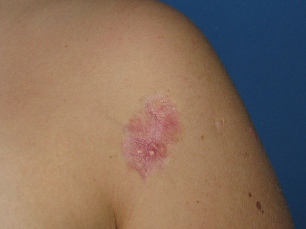
Acosta et al: Intralesional Triamcinolone in Children TABLE 2. Anatomic Location of Keloids Figure 3. The same patient after two treatment sessionswith intralesional triamcinolone.
The median number of injections required per keloid was two (range one to five). The median dosefor each session was 16 mg of triamcinolone acetonide(range 4–40 mg) and the median dose to complete thetreatment was 32 mg (range 4–80 mg).
Analyzing the affected body zone, the average percentage decrease in volume by area was 86% forthe arms, 81% for the chest, 82% for the shoulder, and72% for the auricular area (p < 0.46). The originalvolume diminished by 69% for the keloids resulting Figure 1. Volume comparison before and after therapy.
from varicella, 76% for those after vaccination, and88% for those resulting from trauma or surgical scars(p < 0.16). We observed no correlation between thetime before the start of treatment and response(percentage of reduction) (p < 0.87).
A single infiltration was sufficient in six of the keloids (25%). Generally, two sessions were requiredto achieve the desired effect (range two to five). Threelesions disappeared completely on sonographic exam-ination, and all of them were secondary to wounds.
Central depression at the infiltrated zone was noted inthree patients and was considered a complication ofthe procedure (12.5%). Telangiectasias and fat depos-its appeared in six of the lesions (20.8%). These arewell-described side effects of intralesional steroidinjection, and the patients were advised accordingly(Fig. 4). One keloid did not respond to treatment even Figure 2. A 36-month-old keloid scar secondary to after five infiltrations. It was the only failure in the trauma. Preinjection image.
All patients experienced pain during administra- tion, but only one patient withdrew from the study a decrease of 0.92 cm2 (range 0.05–6.29 cm2), corre- because of pain.
sponding to an 82.7% (0.25–100%) reduction in size Follow-up was 30 months (range 18–53 mos), with between the first and last treatments (p < 0.001) one case of partial recurrence (4%). All patients (Figs. 1–3).


Pediatric Dermatology Vol. 33 No. 1 January/February 2016 with earlobe keloids. The recurrence rate was 4%,which is in agreement with our results and with theresults of the study by Ogawa et al (8). Hamrick'spatients each required at least four injection sessions,which the body area affected could explain; in ourseries, the earlobe was the area that needed the mostinfiltrations and showed the least volume decrease. Inseveral of the studies, the follow-up periods rangedfrom 2 months to 7 years, with a median time ofapproximately 25 months (1,2,9). In our study, themedian follow-up was 30 months (range 18–53 mos).
Although a recurrence could present many years aftertreatment, in our study, the recurrence rate was low inthe short term.
The use of intralesional or topical steroids is one of Figure 4. Fat deposits and telangiectasias.
the most widespread practices in keloid treatment. Itssuccess rate has been reported to range from 50% to100%, with a recurrence rate of 9% to 50% (9). Local side effects include thinning and atrophy of the skin Keloids have been a medical challenge for decades.
and subcutaneous tissues, telangiectasia, fat deposits, The uncertainty of the etiopathogenesis has probably and hypopigmentation (10). Side-effect occurrence prevented effective intervention for avoiding keloid was observed in 20.8% of the cases in our series.
formation. It is difficult to assess the results of therapy Because of low recurrence rate, surgical resection because of the heterogeneity in the study designs followed by radiotherapy is one of regarding race, small series size, the combination of treatments of keloids and is widely used throughout associated therapies, and particularly because of the the world, although there is ongoing fear of the lack of objective measures that ensure comparability development of malignant tumors. Ogawa et al's (10) of results. Many publications define success or failure literature review reported carcinogenesis attributable based on subjective measures such as "good, regular, to the use of radiotherapy for keloids between 1901 and bad." Our study design incorporated the dimen- and 2009. They reported five cases: one in the treated sions of the lesions observed with sonography, which area and four in adjacent tissues. They assert that allows an objective evaluation of the lesion before and radiotherapy is safe with adequate preventive mea- after treatment. Others researchers have used different sures, which are particularly crucial in delicate tissues direct measurements of lesions, including Weshashy such as the neck and mammary glands. Lundell et al and Abdel Hay (4), who used an alginate mold filled (11) conducted a study of thousands of adults with saline solution to estimate the volume more irradiated in childhood for infantile hemangiomas precisely. Sadeghinia and Sadeghinia (5) used a and found a greater incidence of thyroid and mam- caliper to measure the largest dimension of keloid mary gland cancer. Although retinoids have been used width, length, and height.
as an adjunct to surgery, they are not considered first- The injection of triamcinolone acetonide was line therapy. Side effects of long-term systemic shown to be effective in our patients (84.6% volume retinoids in children have been extensively docu- decrease). The analysis of the response to treatment mented and include potential irreversible bone dam- according to the etiology of the keloid and the body age (osteoporosis, ligaments calcification, premature area affected is an important aspect of keloid treat- epiphysis fusion). Although these adverse reactions ment research that has not received sufficient atten- are not related to topical use of retinoids or short tion. Anthony et al (6) treated 256 patients with courses, there are insufficient studies demonstrating intralesional triamcinolone and found differences in efficacy and safety in children younger than 12 years the number of required infiltrations according to the old (2,9). Many studies have examined imiquimod 5% affected area, although their results were expressed cream use in the postoperative period, applying the only in frequency percentage.
medication during a variable period of time of 6 to In the only series conducted exclusively in children, 24 weeks after surgery. Although the recurrence rates Hamrick et al (7) studied the response of excision and were low, nearly all of the reports involved earlobe intralesional triamcinolone acetonide in 15 patients keloids and were small case series. Cacß ao et al (12) Acosta et al: Intralesional Triamcinolone in Children reported the failure of imiquimod 5% cream in the 2. Fruth K, Gouveris H, Kuelkens C et al. Radiofre- prevention of chest keloid recurrence.
quency tissue volume reduction for treatment of auricle Regarding size estimation, the Vancouver scar keloids. Laryngoscope 2011;121:1233–1236.
3. Kim HK, Park MK, Kim BJ et al. The treatment of scale (13) is a good method for evaluating clinical keloids with pneumatic technology: a pilot study. Int J follow-up, but we hypothesize that it is not sufficiently objective to compare results between studies. Nicho- 4. Weshahy AH, Abdel Hay R. Intralesional cryosurgery las et al (14) evaluated the Patient and Observer Scar and intralesional steroid injection: a good combination Assessment Scale in keloids and found that it had no therapy for treatment of keloids and hypertrophic scars.
Dermatol Ther 2012;25:273–276.
pediatric adaptation and that area calculation could 5. Sadeghinia A, Sadeghinia S. Comparison of the efficacy be difficult when assessing nonsurgical scars. The use of intralesional triamcinolone acetonide and 5-fluor- of ultrasound has been attempted, but developing a ouracil tattooing for the treatment of keloids. Dermatol more accurate method is necessary (15).
Our study provides accurate information regarding 6. Anthony ET, Lemonas P, Navsaria HA et al. The cost effectiveness of intralesional steroid therapy for keloids.
keloids, allowing patients and their parents to decide Dermatol Surg 2010;36:1624–1626.
which treatment is best for them, taking into account 7. Hamrick M, Boswell W, Carney D. Successful treat- the keloid characteristics such as location and etiology.
ment of earlobe keloids in the pediatric population. JPediatr Surg 2009;44:286–288.
8. Ogawa R, Huang Ch, Akaishi S et al. Analysis of surgical treatments for earlobe keloids: analysis of 174lesions in 145 patients. Plast Reconstr Surg 2013; Intralesional triamcinolone acetonide therapy for keloids is highly effective in children, without any 9. Gauglitz GG, Korting HC, Pavicic T et al. Hyper- adjuvant method. The average decrease in lesion trophic scarring and keloids: pathomechanisms and volume was 84.6%, and 96% of cases had no current and emerging treatment strategies. Mol Med2011;17:113–125.
recurrence during 18 to 53 months of follow-up.
10. Ogawa R, Yoshitatsu S, Yoshida K et al. Is radiation Complications, seen in 21%, were mild and aesthetic therapy for keloids acceptable? The risk of radiation- in nature and included hypopigmentation, skin atro- induced carcinogenesis. Plast Reconstr Surg 2009;124: phy, and telangiectasia. Our findings indicate a trend toward better response in those lesions caused by a 11. Lundell M, Mattsson A, Hakulinen T et al. Breast cancer after radiotherapy for skin hemangioma in trauma or a surgical scar than in those caused by infancy. Radiat Res 1996;145:225–230.
varicella or vaccination, although this difference was 12. Cacß ao FM, Tanaka V, Messina MC. Failure of not statically significant. Keloids on the earlobe had a imiquimod 5% cream to prevent recurrence of surgi- worse response than those on the arm, chest, or other cally excised trunk keloids. Dermatol Surg 2009;35:629– locations, although the difference was not statistically 13. Sullivan T, Smith J, Kermode J et al. Rating the burn significant. The small size of the sample could have scar. J Burn Care Rehabil 1990;11:256–260.
influenced these results. The duration of the keloid 14. Nicholas RS, Falvey H, Lemonas P et al. Patient- before starting treatment had no effect on the related keloid scar assessment and outcome measures.
treatment result.
Plast Reconstr Surg 2012;129:648–656.
Considering the lack of published data on keloid 15. Aya R, Yamawaki S. Ultrasound elastography to evaluate keloids. Plast Reconstr Surg Glob Open treatment in children, our work contributes by pro- viding evidence of the safety and effectiveness ofintralesional triamcinolone acetonide therapy.
1. Viera MH, Amini S, Valins W et al. Innovative ther- apies in the treatment of keloids and hypertrophic scars.
J Clin Aesthet Dermatol 2010;3:20–26.

Source: http://www.doctoraacosta.cl/wp-content/uploads/2013/08/Triamcinolona-Pediatric-dermatology.pdf

lentech.com.co

Graefes Arch Clin Exp OphthalmolDOI 10.1007/s00417-012-2226-y Visual outcomes and complications following posterioriris-claw aphakic intraocular lens implantation combinedwith penetrating keratoplasty Johannes Gonnermann & Necip Torun &Matthias K. J. Klamann & Anna-Karina B. Maier &Christoph v. Sonnleithner & Antonia M. Joussen &Peter W. Rieck & Eckart Bertelmann Received: 21 August 2012 / Revised: 31 October 2012 / Accepted: 21 November 2012
Promega notes 100: novel biosensors to monitor cellular events in live cells

LIVE-CELL BIOSENSOR Novel Biosensors to Monitor Cellular Events in Live Cells Review of Fan, F. et al. (2008) Novel genetically encoded biosensors using firefly luciferase. ACS Chem. Biol. 3, 346–51. Neal Cosby, Promega Corporation entists targeted the hinge region of the luciferase mol- Drug discovery and life science researchers desire to