Draft consensus guidelines for diagnosis and treatment of shwachmandiamond syndrome

Ann. N.Y. Acad. Sci. ISSN 0077-8923 Draft consensus guidelines for diagnosis and treatment
of Shwachman-Diamond syndrome

Yigal Dror,1 Jean Donadieu,2 Jutta Koglmeier,3 John Dodge,4 Sanna Toiviainen-Salo,5Outi Makitie,5 Elizabeth Kerr,1 Cornelia Zeidler,6 Akiko Shimamura,7 Neil Shah,3Marco Cipolli,8 Taco Kuijpers,9 Peter Durie,1 Johanna Rommens,1 Liesbeth Siderius,10and Johnson M. Liu111 The Hospital For Sick Children, University of Toronto, Ontario, Canada. 2Trousseau Hospital, Paris, France. 3Great OrmondStreet Hospital and Institute of Child Health, London, UK. 4University of Wales Swansea, UK. 5Helsinki University Hospital andChildren's Hospital, University of Helsinki, Helsinki, Finland. 6Hannover Medical School, Hannover, Germany. 7FredHutchinson Cancer Research Center, University of Washington, Seattle, Washington. 8Cystic Fibrosis Center, Ospedale CivileMaggiore, Verona, Italy. 9Emma Children's Hospital, Academic Medical Center, University of Amsterdam, the Netherlands.
10 Youth Health Care, Meppel, the Netherlands. 11The Feinstein Institute for Medical Research, Cohen Children's MedicalCenter of NY, Manhasset and New Hyde Park, NY Address for correspondence: Johnson M. Liu, MD, The Feinstein Institute for Medical Research, Cohen Children's MedicalCenter of NY, Room 255, New Hyde Park, NY 11040, [email protected] Shwachman-Diamond syndrome (SDS) is an autosomal recessive disorder characterized by pancreatic exocrine insuf-
ficiency and bone marrow failure, often associated with neurodevelopmental and skeletal abnormalities. Mutations
in the SBDS
gene have been shown to cause SDS. The purpose of this document is to provide draft guidelines for
diagnosis, evaluation of organ and system abnormalities, and treatment of hematologic, pancreatic, dietary, dental,
skeletal, and neurodevelopmental complications. New recommendations regarding diagnosis and management are
presented, reflecting advances in understanding the genetic basis and clinical manifestations of the disease based on
the consensus of experienced clinicians from Canada, Europe, and the United States. Whenever possible, evidence-
based conclusions are made, but as with other rare diseases, the data on SDS are often anecdotal. The authors welcome
comments from readers.

and spectrum of the human disease. In particular,neurocognitive manifestations such as learning and Management: coordinated care model behavioral disabilities may be under-recognized. Di- Shwachman-Diamond syndrome, first described in versity in how SDS manifests suggests the value of a 1964 (ref [1–3]), is a multi-system disease involv- coordinated multidisciplinary approach to clinical ing the bone marrow, pancreas, bony skeleton, and care. Consensus guidelines presented in this doc- other organs. Decisions about patient management ument aim to improve health care by highlighting are often difficult to make due to the complexity of different aspects of SDS and facilitating early diag- the clinical phenotype, rarity of the disease and the nosis, prevention and therapy.
paucity of large studies. The last report of consensusguidelines for SDS was published in 2002 (ref [4]).
With the identification of the SBDS gene in 2003 (ref General features of SDS [5]), diagnostic criteria have changed. DNA anal- The predominant manifestations of SDS comprise ysis may lead to the diagnosis of SDS before the bone marrow failure, pancreatic exocrine dysfunc- full clinical spectrum is present. Informed clinical tion and skeletal abnormalities.6–8 In addition, the surveillance and the early findings from experimen- liver, kidneys, teeth, brain, and immune system tal models have further highlighted that mutations may also be affected.6,9–13 SDS is also associated in SBDS affect a broad spectrum of functions, which with a propensity for myelodysplastic syndrome has led to a reexamination of the clinical phenotype (MDS) and leukemia.6,9,14–16 SDS is a rare inherited Ann. N.Y. Acad. Sci. 1242 (2011) 40–55 c  2011 New York Academy of Sciences.
Dror et al. Draft consensus guidelines for SDS diagnosis and treatment marrow failure syndrome with an estimated inci- quire pancreatic enzyme supplements as based on dence of 1/76,000 (ref [17]). Although SDS is an evidence of normal fat absorption.9 Although the autosomal recessive disorder, the ratio of males to causative mechanism is unknown, normalization of females reported in the literature with SDS is 1.7 to fat absorption over the years may remain limited to a 1 (ref [10]).
subgroup of patients. Despite the relief in subjectivesymptoms, all patients had a persistent deficit of en- Hematological manifestations. Neutropenia is the
zyme secretion in quantitative studies of pancreatic most common hematological abnormality, occur- ring in nearly all patients. It might be seen in Hepatomegaly is common in young children with the neonatal period,6,18 and it can be either per- SDS. Elevated serum liver enzymes are seen in up sistent or intermittent, fluctuating from severely to 75% of patients, most often in infants and young low to normal levels. In some patients, SDS neu- children, and tend to resolve with age. Although trophils may exhibit defects in migration and there are limited longitudinal data, liver disease ap- pears to have little or no long-term clinical conse- Anemia with low reticulocytes occurs in up to quences.25 Chronic liver disease has not been ob- 80% of the patients. The red blood cells are usu- served in a recent series.26 ally normochromic and normocytic, but can also be Average birth weight is at the 25th percentile.
macrocytic.20 Fetal hemoglobin is elevated in 80% Growth failure with malnutrition is a common fea- of patients.21 The anemia is usually asymptomatic.
ture in the first year of life particularly prior to Thrombocytopenia, with platelets less than 150 × diagnosis. It is attributable to various factors, in- 109/l, is variably seen, as are tri-lineage cytopenias.
cluding inadequate nutrient intake with or without Severe aplasia requiring transfusions has occasion- feeding difficulties, pancreatic insufficiency, and re- ally been reported.6,22,23 current infections.6,10 By the first birthday, over half Bone marrow biopsy usually shows a hypoplas- of patients have dropped below the 3rd percentile tic specimen with increased fat deposition,6,21 but for both height and weight. After diagnosis, and marrows showing normal or even increased cellu- with appropriate therapy, most children show nor- larity have also been observed.10,14 Single-lineage mal growth velocity, but remain consistently below hypoplasia is usually myeloid and occurs in some the 3rd percentile for height and weight.9 patients.9,10 Left-shifted granulopoiesis is a com-mon finding.6,10 Mild dysplastic changes in the Other manifestations. SDS-associated bone dis-
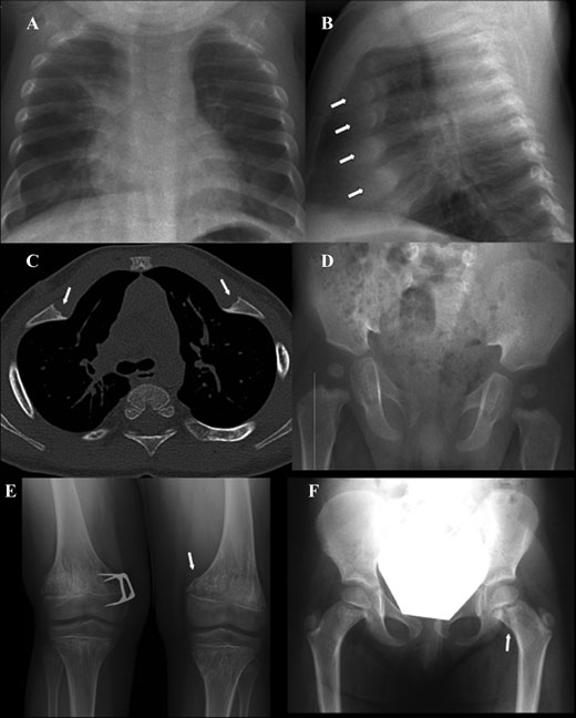
erythroid, myeloid, and megakaryocytic precursors ease includes skeletal dysplasia6,10,27–30 and low- are commonly seen and may fluctuate; however, turnover osteoporosis.31 Skeletal dysplasia usually prominent multilineage dysplasia is less common, presents with metaphyseal changes in the long bones and if it occurs, may signify malignant myeloid and costochondral junctions (Fig. 1), but several other less frequent bone anomalies such as super- Pancreatic dysfunction, nutrition, and liver dis-
numerary fingers and syndactyly have also been de- ease. Variably severe exocrine pancreatic dysfunc-
scribed.12,32 In a small cohort, all had some evidence tion with or without nutrient maldigestion is a hall- of metaphyseal dysplasia at some point, but the fre- mark of SDS.10 Histological specimens of the pan- quency and rate of development are unknown at this creas have revealed extensive fatty replacement of pancreatic acini with preserved islets of Langerhans Delayed dentition of permanent teeth, dental dys- and ductal architecture.3,6 Pancreatic dysfunction is plasia, increased risk of dental caries, and periodon- usually diagnosed within the first six months of life tal disease may also occur. On rare occasions, abnor- and (in 90% of patients) during the first year.9 Duc- malities of the kidneys, eyes, skin, testes, endocrine tular electrolyte and fluid secretion has been shown pancreas, heart, nervous system, and craniofacial to remain normal, but the secretion of proteolytic structures have been reported.6,10,33,34 enzymes is severely decreased leading to steator- How do we diagnose SDS?
rhea.9,24 Spontaneous improvement in pancreaticfunction can occur in later childhood. By 4 years Most patients present in infancy with evidence of of age, almost 50% of patients may no longer re- growth failure, feeding difficulties and/or recurrent Ann. N.Y. Acad. Sci. 1242 (2011) 40–55 c  2011 New York Academy of Sciences.


Draft consensus guidelines for SDS diagnosis and treatment Dror et al. Figure 1. Skeletal radiographic features in SDS. (A and B) Short ribs with marked cupping and widening of the anterior ends
(arrows) in a chest X-ray at 11 months. (C) CT slice shows deformed rib cage with short costae and cupping and irregular widening
of the costochondral junctions (arrows). (D) Broad pelvis, short iliac notches, valgus position of femoral necks and wide proximal
metaphyses of the femora in pelvic X-ray at 11 months. (E) Marked metaphyseal changes with striated bony structure in both hips
and the knees at 14 years. Medial hemiepiphyseodesis was performed on the right distal femur due to genum valgum. A stress
fracture in the left distal femur (arrow). (F) Broad femoral necks with abnormal metaphyseal structure and a stress fracture in the
left femoral neck metaphysis (arrow).

infections. Clinical diagnosis is generally made in Attention should be given to ruling out cystic the first few years of life but occasionally the di- fibrosis (the most common cause of pancreatic in- agnosis may be established in older children and sufficiency) with a sweat chloride test, Pearson dis- even adults. The clinical diagnosis (Table 1) is estab- ease (pancreatic insufficiency and cytopenia, mar- lished by (a) documenting evidence of characteris- row ring sideroblasts and vacuolated erythroid and tic exocrine pancreatic dysfunction and hematolog- myeloid precursors), cartilage hair hypoplasia (diar- ical abnormalities10,35,36 and (b) excluding known rhea and cytopenia, and metaphyseal chondrodys- causes of exocrine pancreatic dysfunction and bone plasia, and more common in certain isolated pop- marrow failure.
ulations such as the Amish), and other inherited Ann. N.Y. Acad. Sci. 1242 (2011) 40–55 c  2011 New York Academy of Sciences.
Dror et al. Draft consensus guidelines for SDS diagnosis and treatment Table 1. Clinical and molecular diagnostic criteria
Diagnostic criteriaClinical diagnosis: Fulfill the combined presence of hematological cytopenia of any given lineage (most often neutropenia) and exocrine pancreas dysfunction Hematologic abnormalities may include: a. Neutropenia <1.5 x 10⁹/L on at least 2 occasions over at least 3 monthsb. Hypoproductive cytopenia detected on 2 occasions over at least 3 months Tests that support the diagnosis but require corroboration: a. Persistent elevation of hemoglobin F (on at least 2 occasions over at least 3 months apart)b. Persistent red blood cell macrocytosis (on at least 2 occasions over at least 3 months apart), not caused by other etiologies such as hemolysis or a nutritional deficiency Pancreatic dysfunction may be diagnosed by the following: a. Reduced levels of pancreatic enzymes adjusted to age [fecal elastase, serum trypsinogen, serum (iso)amylase, serum lipase] Tests that support the diagnosis but require corroboration: a. Abnormal 72 hr fecal fat analysisb. Reduced levels of at least 2 fat-soluble vitamins (A, D, E, K)c. Evidence of pancreatic lipomatosis (e.g. ultrasound, CT, MRI, or pathological examination of the pancreas by autopsy) Additional supportive evidence of SDS may arise from: a. Bone abnormalitiesb. Behavioral problemsc. Presence of a first degree-family member diagnosed before with SDS Other causes pancreatic insufficiency should be excluded, in particular when the SBDS gene mutation analysis Molecular diagnosis: biallelic SBDS gene mutation
Positive genetic testing for SBDS mutations known or predicted to be deleterious, e.g. from protein modeling or expression systems for mutant SBDS Caveats:Many situations arise when molecular diagnosis is NOT confirmatory in the presence of clinical symptoms:No identified mutations (about 10% of cases)Mutation on one allele onlyGene sequence variations that have unknown or NO phenotypic consequence:A novel mutation, such as a predicted missense alteration, for which it is not yet possible to predict whether it isdisease-causing.
SBDS polymorphisms on one or both alleles. Large population studies may be needed to exclude a sequence polymor-phism as a bona fide irrelevant variant.
bone marrow failure syndromes (such as dyskerato- atic reserve loss of 98% must occur before signs sis congenita).
and symptoms of maldigestion are present. Thus,72-hour fecal fat balance studies may be normal de- Exocrine pancreatic phenotype spite a significant defect in pancreatic acinar func- The clinical diagnosis of the pancreatic phenotype tion. The terms pancreatic insufficiency (PI) and is challenging as most pancreatic function tests lack pancreatic sufficiency (PS) have been coined to dis- sufficient sensitivity and/or specificity. This is com- criminate between subjects with PI, who require plicated by the fact that nearly half of subjects pancreatic enzymes supplements with meals and with SDS show improvement in exocrine pancre- those with PS, who invariably have loss of pancreatic atic function with advancing age. Exocrine pancre- reserve but lack clinical evidence of maldigestion.
Ann. N.Y. Acad. Sci. 1242 (2011) 40–55 c  2011 New York Academy of Sciences.
Draft consensus guidelines for SDS diagnosis and treatment Dror et al. For these reasons, alternative approaches are rec- perfusion technique to quantify timed collections ommended to assess patients with a suspected di- of pancreatic juice during hormonal stimulation agnosis of SDS for evidence of pancreatic dysfunc- with cholecystokinin and secretin provided use- tion. Serum pancreatic enzyme concentrations are ful information concerning the pathophysiology of useful markers of the pancreatic phenotype in pa- the exocrine pancreas. However, this complex, in- tients with SDS.37 Serum immunoreactive trypsino- vasive test has little role in a clinical setting and gen concentrations are low (<6 ␮g/L) in patients is largely used only in research studies. Alternative with SDS who have PI. However, in patients with PS, non-quantitative methods of collecting secretions, serum trypsinogen concentrations are usually above including aspiration of pancreatic juice with a duo- 6 ␮g/L, and in one fifth of PS patients, measured denoscope or single lumen duodenal tube are not concentrations are within the reference range. Thus, recommended because they show considerable test a low serum trypsinogen is helpful in identifying the variability and approximately 25% of PS subjects pancreatic phenotype, but a normal value does not with low pancreatic reserve may be misclassified as exclude impaired exocrine pancreatic function. In contrast, serum pancreatic isoamylase activities in Hematologic phenotype SDS patients are uniformly low at all ages, regard- The hematologic phenotype is most frequently char- less of pancreatic status or trypsinogen concentra- acterized by intermittent or persistent neutropenia, tion. Unfortunately, serum isoamylase activity can- but cytopenias of other blood cell lineages are fre- not be used as a sole marker of the SDS pancreatic quently present. Red blood cell macrocytosis, high phenotype because isoamylase production shows hemoglobin F, and varying degrees of marrow hy- age-dependent postnatal development. Healthy in- poplasia are also typical findings.
fants have low pancreatic isoamylase concentrations Chromosome breakage studies with diepoxybu- (similar to those observed in SDS), which rise and tane or mitomycin C are recommended to exclude achieve adult values by approximately three years of Fanconi anemia, unless the history, physical exam- ination and initial work-up are diagnostic for SDS.
To overcome these limitations, serum trypsino- Bone marrow aspiration and biopsy are essential gen, isoamylase, and age have been incorporated for initial evaluation and should include assessment into a diagnostic rule for the SDS pancreatic phe- of cellularity, differential, iron stain and cytogenet- notype, using the Classification and Regression Tree ics. Bone marrow cytogenetic finding of i(7q) or (CART) analysis of Breiman et al.37 With the excep- del(20q) is highly associated with SDS. Virology
tion of patients less than 3 years of age, the diagnos- studies (e.g. Epstein–Barr virus, cytomegalovirus, tic rule effectively distinguished control individuals and B19 parvovirus) may be pursued as clinically from patients with a confirmed clinical diagnosis of indicated to exclude other causes of bone marrow suppression and a failure to thrive.
Several alternative non-invasive approaches to es- tablish or exclude pancreatic dysfunction may be Skeletal phenotype considered, including multi-dimensional imaging When present in association with hematologic (ultrasound, CT, or MRI) for evidence of fatty re- or pancreatic abnormalities, characteristic skele- placement of the pancreas, and fecal enzyme con- tal abnormalities are strongly suggestive of SDS.
centrations of pancreatic elastase or chymotrypsin.
SDS bone dysplasia is characterized by short Concentrations of fecal elastase less than 200 ␮g/g stature, delayed appearance but subsequent nor- stool offer evidence of severe pancreatic dysfunc- mal development of secondary ossification cen- tion, and a fecal elastase <100 ␮g/g is suggestive ters, and by variable metaphyseal widening and of maldigestion due to exocrine pancreatic insuf- irregularity that is most often seen in the ribs ficiency. Fecal fat balance studies provide direct in early childhood and in the proximal and dis- evidence of the severity of malabsorption, but as tal femora later in childhood and adolescence.10,27 mentioned above, they do not indicate a specifically Rarely, skeletal involvement may be extremely se- pancreatic cause if fat malabsorption is found.
vere with generalized bone abnormalities.38 Al- The "gold standard" method of directly measur- though metaphyseal changes often become unde- ing pancreatic secretion using an intestinal marker tectable and clinically insignificant over time, they Ann. N.Y. Acad. Sci. 1242 (2011) 40–55 c  2011 New York Academy of Sciences.
Dror et al. Draft consensus guidelines for SDS diagnosis and treatment may also progress and result in limb deformities, as severe marrow failure, myelodysplastic syndrome most commonly at the hips and the knees, or stress (MDS), or leukemia.
fractures of the femoral necks (Fig. 1).27 In addition When infections regularly recur, immunoglob- to metaphyseal chondrodysplasia, SDS associates ulin levels and post-vaccination antibodies should with early-onset low-turnover osteoporosis char- be screened to exclude an associated immunodefi- acterized by low bone mass and vertebral fragility Systematic evaluation of neutrophil chemotaxis is not considered a necessity in the usual follow up Other clinical findings of patients.
Short stature with or without malnutrition is also acommon feature of SDS. Hepatomegaly with mild to moderate biochemical abnormalities of the liver Once the diagnosis of SDS is suspected or estab- are common findings in infants and young children lished, objective testing for assessment of pancreatic function status is recommended. To determine PSor PI status, serum trypsinogen concentration offers Molecular testing useful screening information: As the clinical diagnosis of SDS is usually difficult (a)If values are undetectable or low, a 72-hour fat and patients may present at a stage when no clinical balance study may be done to confirm PI status.
pancreatic insufficiency is evident, it is advisable Since most newly diagnosed subjects are infants to test most or all suspected cases for mutations or children, careful documentation of ingested in the SBDS gene (Table 1). It is noteworthy that fat (and other macronutrients) will enable de- about 10% of the SDS patients may be negative for termination of coefficient of fat absorption as mutations, and that de novo SBDS mutations have well as provide insight into total calorie intake.
been identified in some families.
(b)If values are 6 ␮g/L or above, PS status should be confirmed by 72-hour fat balance study as How to monitor a patient after a diagnosis
described. Recent studies in patients with cys- tic fibrosis have, however, shown that duplicate Recommended baseline testing are listed in Table 2.
measurements of the coefficient of fat absorp-tion often show wide variation.
(c)Measurement of fecal elastase or chymotrypsin Hematological evaluation should include complete is widely used in Europe as an alternative indi- blood count (CBC), mean corpuscular volume, cator of pancreatic insufficiency, although it has peripheral blood smear, differential, reticulocyte not been validated in a large series of SDS pa- count, fetal hemoglobin level and coagulation tests tients. It has the theoretical advantage of being a in case of clinical bleeding symptoms. If the diagno- specific test of pancreatic function, whereas fat sis of SDS is suspected or confirmed, bone marrow absorption can of course be abnormal in non- aspirate smear, biopsy, and cytogenetic evaluation pancreatic disorders such as celiac disease.
is recommended as a baseline examination (see sec- Baseline fat soluble vitamin levels (A, D, E) and tion IV for further discussion).
prothrombin time, as a surrogate marker for vita- Complete blood count is a basic parameter that min K status, should be done. Low values should be needs to be monitored: CBCs should be consid- correlated with results of pancreatic function test- ered every 3–6 months in stable patients. Any clin- ing and in patients with PI, should be repeated ap- ical complications, including recurrent infections, proximately one month after instituting enzyme re- bruising, asthenia or pallor may require a CBC be- placement therapy. Persistently low levels in the face tween scheduled examinations. The purpose of the of good compliance with enzyme therapy will re- routine CBC is to determine the baseline profile of quire fat-soluble vitamin supplements. Fat-soluble the patients, to assess the risk for infections and vitamins should be monitored on at least a yearly possibly to detect particular features related to nu- basis, and may include (vitamin K-dependent) co- tritional deficits, such as iron or folate deficiency agulation parameters when clinical symptoms are and to detect evolving marrow abnormalities such Ann. N.Y. Acad. Sci. 1242 (2011) 40–55 c  2011 New York Academy of Sciences.
Draft consensus guidelines for SDS diagnosis and treatment Dror et al. Table 2. Clinical tests at diagnosis and at follow-up
At Diagnosis
At Follow-up
SBDS gene mutation (test may Yes, if not done at diagnosis be offered to family memberhematopoietic stem celltransplant donors) Genetic counselling (molecular test may beoffered to family membersfor screening of carriers) Hematology and immunology 2–4 times / year Bone marrow aspirate and Every 1 to 3 years or as clinically indicated Fe, folate, B12 levels As clinically indicated IgG, IgA, IgM levels Post vaccination serology As clinically indicated Lymphocyte phenotype As clinically indicated As clinically indicated Pancreatic enzymes (choice based on local availability:serum trypsinogen,isoamylase, 72-hour fatbalance test, elastase, etc.) Fat-soluble vitamins A, D, E, 1 mo after pancreatic enzyme therapy, then 1-2 times/ and prothrombin time (surrogate for vitamin K) Other vitamins and As clinically indicated Liver biochemistry panel As clinically indicated Pancreatic imaging Skeletal system, growth Growth evaluation: height, Yearly at follow-up weight and headcircumference As clinically indicated Baseline study: once during prepuberty Follow-up study: once during puberty, then as clinically indicated Oral and dental care Once per year and when clinically indicated Ann. N.Y. Acad. Sci. 1242 (2011) 40–55 c  2011 New York Academy of Sciences.
Dror et al. Draft consensus guidelines for SDS diagnosis and treatment Table 2. Continued
At Diagnosis
At Follow-up
Standardized developmental screening measure: Infancy/-preschool age Neuropsychological assessment of domains: At ages 6–8, 11–13, 15–17 Intellectual abilitiesAttention including working memory,sustained attention and divided/dual attentionHigher order languageVisual-motor integration and speedExecutive functioningAcademic achievementBehaviour (self report and parent proxy)Adaptive Functioning (parent proxy) There are no published guidelines on dosing of ists (e.g., speech and language therapy, occupational pancreatic enzyme supplements in SDS patients therapy, developmental pediatrician, developmen- with PI. Furthermore, there are few published data tal psychologist) as needed. Serial neuropsycholog- demonstrating efficacy of enzyme replacement ther- ical assessments are indicated, at minimum, when a apy. For this reason, published treatment guidelines child is approximately 6, 12, and 15 years of age to for subjects with cystic fibrosis may be considered.39 correspond with brain development and changes inexpectations at school.
Nutritional statusNewly diagnosed infants with SDS are commonly malnourished. Therefore, careful baseline assess-ment of height and weight and anthropometric Definition of hematological complications measures are recommended. Once appropriate ther- While neutropenia (even severe) is a typical feature apy is introduced, malnutrition should be corrected of SDS, anemia (<7 g/dl or 4.3 mmol/L or if symp- by one year of age.
tomatic) and thrombocytopenia (<20 × 109/L orif symptomatic) are additional complications that require prompt evaluation and medical decision.
Skeletal survey is recommended at the time of the Classification of the different forms of marrow diagnosis. The follow-up is based on individual clin- failure in SDS is complex and poorly understood. In ical and radiological findings. For biochemical as- general, cytogenetic studies should be performed sessment and bone mineral density evaluation, see concurrently with morphology studies. Aplastic section on bone abnormalities.
anemia (hypoproliferative cytopenia without dys- plastic morphology and usually without clonal evo-lution) and myelodysplastic syndrome (cytopenia Annual reviews—ideally by a dentist experienced with dysplastic morphology and clonal evolution) in orthodontic approaches and/or periodontal represent the two main categories of complications.
disease—are generally recommended.
However, most of the common scenarios seen in SDS differ from the standard definitions estab- A characteristic pattern of learning and behav- lished by World Health Organization (WHO) crite- ioral difficulties is common in SDS.40 It is there- ria,41 because the bone marrow morphology from fore important to monitor and support neurode- SDS patients often bears mild dysplastic changes velopment. Standardized developmental checklists in the erythroid, myeloid and megakaryocytic se- should be used routinely to assess infant, toddler and ries, even in the absence of clonal cytogenetic preschooler development with referrals to special- Ann. N.Y. Acad. Sci. 1242 (2011) 40–55 c  2011 New York Academy of Sciences.
Draft consensus guidelines for SDS diagnosis and treatment Dror et al. erature review47 reveals that subjects with SDS Aplastic anemia can be divided into moderate and commonly show clonal marrow cytogenetic ab- severe subcategories.42,43 Severe disease is defined normalities (CMCA), MDS or AML. Among those by depression in two of three blood counts (retic- identified with CMCA/MDS in childhood, approx- ulocytes <40,000/␮L, platelets <20,000/␮L, neu- imately 50% progressed to overt leukemia over a trophils <500/␮L) in the presence of a hypocellular range of 1 to 37 years. Remarkably, males constituted bone marrow biopsy (<25% cellularity or <50% 68% and 92% of all subjects with CMCA/MDS and cellularity and <30% hematopoietic cells) without significant fibrosis. Moderate disease is defined as The bone marrow cytogenetic abnormalities failure to meet the criteria for severe disease but with i(7q) and del(20q) are quite common in SDS, occur at least two diminished blood counts (reticulocytes less frequently in other malignancies or marrow fail- <40,000/␮L, platelets <40,000/␮L, neutrophils ure syndromes, and can regress spontaneously.16,48 <l,500/␮L) with a hypocellular bone marrow These specific cytogenetic changes may be relativelybiopsy.
specific for SDS and, in isolation, may not be an The diagnosis of aplastic anemia is usually, but absolute harbinger of malignancy. In general, cyto- not always, considered in the absence of clonal mar- genetic abnormalities of unclear clinical significance row cytogenetic abnormalities (CMCA). Aplastic should be interpreted in the context of the marrow anemia may be transient (lasting less than 3 months) morphology and blast count.14,15,48–54 Of these pa- or may persist past 3 months, becoming clinically tients, some developed severe aplasia, while others significant (J. Donadieu, unpublished data).
progressed to more severe MDS/AML. SDS patientsmay also present with MDS at the stage of refrac- Clonal marrow cytogenetic abnormality tory cytopenia with dysplasia14,15,48–54 or with excess Clonal marrow cytogenetic abnormality (CMCA) blasts, some of whom progress to AML.
is defined by: two or more bone marrow cells (out Various types of AML have been described in SDS of twenty) with gain of the same chromosome or patients: AML-M0, M2, M4, M5, and M6. Acute cytogenetic abnormality or three or more cells with lymphoblastic leukemia and juvenile myelomono- loss of the same chromosome, as detected by G- cytic leukemia were rare. AML-M6 was particu- banding; or a cytogenetic abnormality detected by larly common in SDS, occurring in about 30% of fluorescence in situ hybridization (FISH) analysis in cases with classifiable leukemia. Malignant myeloid higher frequency than the reference values of the transformation into MDS and AML in SDS patients lab, as well as higher than in the concurrently tested while on G-CSF therapy has been reported,49,55,56 control sample.
but the causal relationship is unproven. SDS-related Diagnostic criteria for MDS and AML leukemia carries a poor prognosis if treated with The critical component for MDS is dysplastic mor- chemotherapy alone. However, due to the improv- phology, as defined by the WHO.41 Published cri- ing outcome of stem cell transplantation in patients teria for MDS in children include two out of the over the past years, the prognosis of SDS with sec- following three items: chronic trilineage cytope- ondary leukemia has improved accordingly, but data nia, prominent bi-lineage cytopenia, clonal marrow are still limited.
cytogenetic abnormality, marrow myeloblast countbetween 5–29%.44,45 However, since cytogenetic abnormalities as well as mild dysplastic features In cases presenting with severe pancytopenia, bone occur in some SDS patients without progression marrow aspirate, biopsy, and cytogenetic examina- to AML, the markers that discriminate MDS from tion are mandatory. However, the indications for the aplastic phase are still debatable. AML is de- routine bone marrow smear and bone marrow cy- fined by a marrow myeloblast count of ≥20% togenetics are controversial. To date, in the absence (WHO)41 or ≥30% (French American British of severe cytopenia, bone marrow cytogenetic anal- ysis has not generally been predictive of outcome.
There are two current classification systems for However, non-i(7q) abnormalities of chromosome pediatric MDS,44,45 but the prognostic significance 7, particularly monosomy 7, are associated with of the systems has not yet been studied. A lit- poor outcomes and may present with advanced Ann. N.Y. Acad. Sci. 1242 (2011) 40–55 c  2011 New York Academy of Sciences.
Dror et al. Draft consensus guidelines for SDS diagnosis and treatment MDS/AML or progress from earlier stages of MDS.
bone marrow failure, and for whom an HSCT donor In addition, systematic bone marrow cytogenetic is unavailable.
examination may have a role in surveillance in pa-tients receiving long-term therapy with granulocyte Prevention and treatment of infections colony-stimulating factor (G-CSF, see below).
Patients with acute infectious episodes, suggested In summary, bone marrow aspirate and biopsy by fever or any acute symptoms need to be evalu- are recommended at the time of diagnosis of SDS, in ated urgently. Some patients can be treated with oral cases of CBC changes, and annually in patients who antibiotics, while patients with severe neutropenia are treated with G-CSF therapy. In a patient with sta- or those suspected to have severe infections should ble clinical status and complete blood counts (not be hospitalized and treated with intravenous antibi- on G-CSF), a bone marrow aspirate with cytoge- otics with broad-spectrum coverage until improve- netic examination can be proposed routinely every ment. G-CSF treatment should also be considered 1–3 years.
during infections in patients with severe neutrope-nia. In cases of recurrent infections or severe chronic Treatment of hematologic and infectious
stomatitis with profound neutropenia, long-term G-CSF therapy may be considered (see above).
Bleeding episodes Thrombocytopenia and anemia may require respec- In the presence of thrombocytopenia or low vita- tive chronic transfusions, with institution of an iron- min K-dependent coagulation factors, bleeding may chelation program as clinically indicated. If trans- occur. Mild to moderate bleeding episodes can be fusions are indicated, blood products need to be treated with local measures (xylometazoline 0.05% nose spray), tranexamic acid, or aminocaproic acid.
Granulocyte colony stimulating factor When coagulation is affected by low vitamin K The majority of patients do not need granulocyte and/or, rarely, abnormal liver function, vitamin K colony stimulating factor (G-CSF) due to the low in- should be administered. Platelet transfusions are cidence of infections. Chronic use of G-CSF should indicated in an SDS patient with severe bleeding be considered for recurrent invasive bacterial and/or and thrombocytopenia. Prophylactic administra- fungal infections in the presence of severe neu- tion of platelets should be considered for patients tropenia. G-CSF given for profound and persistent with platelet counts of <10 × 109/L or for those neutropenia has been effective in inducing a clini- with a known tendency to have significant bleeding cally beneficial neutrophil response. Patients may re- spond to an intermittent schedule with low doses of For surgery or invasive procedures, platelets G-CSF (e.g. 2–3 ␮g/kg every 3 days) or may require should be transfused as clinically indicated. When higher doses continuously. The aim of long-term known or suspected coagulation defects are present, G-CSF treatment is not to obtain normal hema- infusion of fresh frozen plasma or plasma-derived tological parameters but to prevent infections. In coagulation products (such as prothrombin com- cases of G-CSF resistance, associated with severe plex, containing factors II, VII, IX, and X) may be infections, hematopoietic stem cell transplantation (HSCT) should be considered.
Female patients suffering from blood loss dur- ing menstruation may benefit from pharmacologic treatment to induce amenorrhea.
Data are scarce regarding response rates to andro-gens in SDS patients. A few patients have received MDS and AML: chemotherapy androgens, and responses have been reported. How- In MDS secondary to SDS, standard chemotherapy ever, androgens are generally not recommended as regimens are not indicated and an attempt should first line therapy for severe bone marrow failure in be made to provide HSCT on an urgent basis. High SDS. Underlying liver abnormalities seen in SDS dose chemotherapy is therefore mainly indicated for may lead to higher liver toxicity than that seen in conditioning prior to HSCT.
Fanconi anemia. The use of androgens should prob- Standard chemotherapy for AML can be effec- ably be reserved for patients who do not have severe tive to temporarily control the disease. However, Ann. N.Y. Acad. Sci. 1242 (2011) 40–55 c  2011 New York Academy of Sciences.
Draft consensus guidelines for SDS diagnosis and treatment Dror et al. chemotherapy alone has been unsuccessful in ob- the second or third decade (younger patients gen- taining a prolonged complete remission in SDS.
erally have better outcomes following HSCT). Most Therefore, due to a high risk of persistent aplasia, data have been collected over the past 20 years, and an urgent search for a related or unrelated donor for current results may be more promising due to bet- HSCT should be initiated and minimal chemother- ter standards for donor searches and treatment of apy to provide interim disease control should be Complications from chemotherapy or HSCT are more common in SDS patients than in pa- Hematopoietic stem cell transplantation tients with idiopathic blood dyscrasias. In a re- Indications for HSCT. The criteria for considering
view of 36 patients with SDS who had been patients for HSCT (related or alternative) include: <7 g/L or with HSCT with or without irradiation, 83% (4.3mmol/L), absolute neutrophil count < died from complications related to the therapy, in- 0.5 × 10⁹/L with recurrent infections, platelet cluding prolonged severe aplasia, infections, car- count <20 × 109/L] diotoxicity, neurological and renal complications, (b) MDS with excess blasts veno-occlusive disease, pulmonary disease, post- (c) Overt leukemia transplant graft failure, and GVHD. Toxicity, par- In cases of frank leukemia, the patient may be ticularly cardiac toxicity,64 seems more frequent if started on chemotherapy to reduce tumor load be- the indication is MDS/acute leukemia rather than fore HSCT, but an effort to find a donor should be aplastic anemia. Recently, an attenuated condition- made at the time of diagnosis because of the high ing regimen has been proposed in order to limit risk of therapy-related aplasia.
In considering the indications for HSCT, one should also allow for the possibility of spontaneous Treatment of pancreatic dysfunction,
recovery from aplasia. Depending on the level of nutrition and liver disease
potential immediate risks of the severe cytopenia, a Pancreatic enzymes monitoring period of up to 3 months can be con- The clinical response to enzyme treatment in pa- sidered while concurrently initiating the process of tients with SDS, in contrast to patients with cystic HLA typing and donor search.
fibrosis for whom there may be additional intesti- Conditioning regimen and GVHD prophylaxis.
nal factors, is usually excellent, although growth At present, HSCT provides the only curative option may continue to be restricted for skeletal reasons.
for the hematological complications in SDS. Re- The natural history of SDS suggests that pancre- ported cases of SDS patients who have undergone atic function may improve to sufficient levels in HSCT include no more than 80 patients world- many patients to allow them to discontinue en- wide.57,58 Many different conditioning/supportive zyme supplementation as they become older. The regimens in small groups of patients render general pancreatic status of all patients should therefore be conclusions and recommendations difficult. Glob- reassessed from time to time, according to their clin- ally, it appears that the results depend on the type ical progress.
of donor (genotypically identical donor transplants Once the diagnosis is made, and steator- better than matched unrelated donor or MUD rhea confirmed, pancreatic enzyme replacement transplants) in almost all reports. However, the in- should be started. The initial dose should be dications for HSCT also appear to be a clear deter- 2,000 lipase units/Kg body weight/day. The dos- minant of survival. The survival of patients receiv- ing guidelines for subjects with cystic fibrosis ing a transplant for aplastic anemia is about 80%, disease (maximum 10,000 lipase units/kg body while the survival of patients receiving a transplant weight/day) should be followed.39 Pancreatin is for MDS or acute leukemia remains between 30 and taken with all meals and snacks that con- 40%. This disparity is likely due in part to differences tain protein, fat or complex carbohydrates. In in the ages of recipients, because aplastic anemia is children with persistent fat malabsorption despite usually a complication in the first decade of life, optimal dose of replacement, an H2-receptor an- whereas MDS/AML is more likely a complication of tagonist or proton pump inhibitor may be given Ann. N.Y. Acad. Sci. 1242 (2011) 40–55 c  2011 New York Academy of Sciences.
Dror et al. Draft consensus guidelines for SDS diagnosis and treatment in addition. Higher requirements of pancreatic en- tal surface can lead to decay and can be severe in zymes should alert the clinician to the possibility of some cases. Gastric acid reflux can lead to tooth a concomitant unrelated enteropathy.
surface loss or erosion. Regular dental care and ap- Enteric-coated enzyme preparations prevent gas- propriate advice from an early age are crucial to tric acid-peptic degradation and therefore deliver minimize these oral and dental problems.
a higher concentration of enzymes to the intestine Treatment of bone abnormalities
than uncoated preparations. The capsules shouldbe swallowed whole, without chewing. If the patient Treatment and follow-up cannot swallow capsules, they can be opened and the Bone deformities due to metaphyseal chondrodys- enteric-coated granules mixed with milk, juice or plasia, usually located at the hips or the knees, may pureed fruit. The resulting mixture should be swal- require orthopedic consultation and surgical in- lowed immediately without chewing. Pancreatin is terventions. Low-turnover osteoporosis may result inactivated at high temperatures, and excessive heat from a primary defect in bone metabolism that is should be avoided when the granules are mixed with related to the bone marrow dysfunction and neu- liquids or food.
tropenia. Efforts should be made to optimize generalpreventive measures such as nutrition and intake of Vitamin supplements fat-soluble vitamins, as well as to promote weight- Blood levels of fat-soluble vitamins should be mea- bearing exercise. Supplementation with vitamin D sured every 6 to 12 months in young children, and (in addition to other fat-soluble vitamins) and cal- supplementary therapy started if values are low. It cium should be commenced if dietary intakes are not is important to ensure compliance with pancreatic sufficient. It is presently unknown whether bispho- enzyme supplementation, as deficiencies of these vi- sphonates, anti-resorptive agents used to treat post- tamins are an indirect marker of fat malabsorption.
menopausal high-turnover osteoporosis, are safe Dietary advice and surveillance and efficacious in SDS osteoporosis. Optimal treat- Height and weight should be documented at every ment for SDS osteoporosis remains to be estab- clinic visit. All patients should receive an evaluation by a dietitian. Poor appetite and behavioral feeding Radiography and bone densitometry. Assessment
difficulties are common. Such children should have of bone dysplasia (Tables 2 and 3): at diagnosis, ra- a careful psychology assessment and support offered diographic skeletal survey; follow-up based on in- to the family by a clinical psychologist.
dividual clinical and radiographic findings, X-rays If oral intake is suboptimal nutritional supple- for detection of deformities or stress fractures (hips, ments should be considered. If there are ongoing knees). Assessment of osteoporosis: bone densitom- concerns about poor weight gain despite adequate etry by DXA, at prepuberty (baseline study), during pancreatic enzyme replacement therapy, it may be pubertal years, postpubertal follow-up studies based necessary to assess the child for other causes or con- on individual findings (low BMD, vertebral com- ditions such as gastro-esophageal reflux, food al- pressions, multiple peripheral fractures). Caution lergy and enteropathy.67 should be exercised when interpreting DXA results In severe cases of persistent failure to thrive or in patients with SDS; small body size and delayed feeding difficulties, as a last resort a gastrostomy pubertal development affect BMD results.
insertion can be considered to allow overnight feed-ing, but weaning should be attempted once the pa- tient is stable.
plasma parathyroid hormone (PTH) should bemonitored as part of routine follow-up and main- Treatment of dental complications
tained within normal limits after the diagnosis.
Oral and dental problems are common in children with SDS68. Ulceration of the oral mucosa can be as- and support
sociated with neutropenia. The frequency and sever-ity of the ulceration is variable. Enamel defects have Deficits in cognitive abilities across numerous do- been noted, in both the deciduous and permanent mains of functioning are evident in the majority of dentitions. Areas of faulty mineralization of the den- individuals with SDS at varying levels of severity Ann. N.Y. Acad. Sci. 1242 (2011) 40–55 c  2011 New York Academy of Sciences.
Draft consensus guidelines for SDS diagnosis and treatment Dror et al. Table 3. Longitudinal changes in skeletal phenotype in SDS
Infancy and early childhood Delayed appearance of secondary ossification centers Wrist, hand, femur Wide, irregular metaphyses Osteopenia, Wormian bones Tubular bones, skull Slow development of secondary ossification centers Wrist, hand, femur Irregularity and sclerosis of metaphyses Tubular bones, spine Late childhood/ puberty Irregularity, sclerosis and asymmetrical growth of metaphyses Stress fractures, deformity Compression fractures Compression fractures indicating heterogeneity. Parental report indicates ioral, social, and adaptive functioning are war- that over 50% of children experience delayed lan- ranted from time of diagnosis through to adult- guage development.6,40 Below average intellectual hood. Specifically, during the infancy/pre-school reasoning abilities are also evident6,40,69,70 with ap- period (diagnosis to 4 years of age), it is ad- proximately 1 in 5 meeting the diagnostic criteria for vised that comprehensive developmental check- an intellectual disability (i.e., IQ < 2nd percentile).40 lists be used so that referrals to specialists (i.e., Difficulties in visual reasoning and visual-motor speech and language therapist, occupational ther- integration,40,70 higher order language functioning apist, developmental pediatrician, developmental (e.g. understanding figurative expressions, knowl- psychologist), assessment and intervention can oc- edge of synonyms), executive problem solving and cur at the earliest sign of possible issues. In ad- attention have also been documented.40 dition, it is recommended that serial neuropsy- Significant behavioral issues are commonly re- chological assessments be completed to coincide ported. In a study of 32 children / adolescents (ages with key stages of brain maturation, namely 6–8, 6 through 17),40 19 percent had prior diagnosis of at- 11–13, and 15–17 years of age. These age groups tention deficit hyperactivity disorder, pervasive de- also parallel changes in expectations in learn- velopmental disorder or oppositional defiant disor- ing at school. Assessments should include eval- der while an additional 31 percent were reported to uation of intellectual abilities, attention (work- have some combination of inattention, restless, im- ing memory, sustained attention, and divided/dual pulsivity, and oppositional behavior. In addition, on attention), higher order language, visual percep- behavioral rating scales, parents indicated a height- tion, visual-motor functioning, executive skills, aca- ened frequency of attention problems (50%) and demic readiness/achievement, behavior, and func- social problems (34%). The neurocognitive deficits tional independence. The identification of an have been found to be independent of pancreatic individual's strengths and weaknesses, consequently involvement, otitis media, having a chronic illness, leads to individualize recommendations for inter- family environment, and age.40 Given the structural vention, which are reviewed and adapted at the abnormalities that are evident on neuro-imaging of follow-up assessment at the next critical stage of de- the brain,71–73 neurocognitive and neurobehavioral velopment. Counselling for parents should parallel issues are likely the consequences of SBDS gene dys- the neuropsychological assessments of their child to function on the brain.
support them in enhancing interactions with, and indeveloping realistic expectations for, their child.
Assessment, monitoring, and treatmentIn order to maximize ongoing development, com- Conflicts of interest
prehensive assessments using standardized tests andclinical observation to monitor cognitive, behav- The authors declare no conflicts of interest.
Ann. N.Y. Acad. Sci. 1242 (2011) 40–55 c  2011 New York Academy of Sciences.
Dror et al. Draft consensus guidelines for SDS diagnosis and treatment 19. Rothbaum, R.J., D.A. Williams & C.C. Daugherty. 1982.
Unusual surface distribution of concanavalin A reflects a 1. Shwachman, H., L.K. Diamond, F.A. Oski & K.T. Khaw.
cytoskeletal defect in neutrophils in Shwachman's syndrome.
1964. The syndrome of pancreatic insufficiency and bone Lancet 2: 800–801.
marrow dysfunction. J. Pediatr. 65: 645–663.
20. Woods, W.G., W. Krivit, B.H. Lubin & N.K. Ramsay. 1981.
2. Nezelof, C. & M. Watchi. 1961. [Lipomatous congenital hy- Aplastic anemia associated with the Shwachman syndrome.
poplasia of the exocrine pancreas in children. (2 cases and In vivo and in vitro observations. Am. J. Pediatr. Hematol. review of the literature)]. Arch. Fr. Pediatr. 18: 1135–1172.
Oncol. 3: 347–351.
3. Bodian, M., W. Sheldon & R. Lightwood. 1964. Congenital 21. Dror, Y. & M.H. Freedman. 1999. Shwachman-Diamond Hypoplasia of the exocrine pancreas. Acta. Paediatr. 53: 282–
syndrome: An inherited preleukemic bone marrow failure disorder with aberrant hematopoietic progenitors and faulty 4. Rothbaum, R., J. Perrault, A. Vlachos, et al. 2002.
marrow microenvironment. Blood 94: 3048–3054.
Shwachman-Diamond syndrome: report from an interna- 22. Tsai, P.H., I. Sahdev, A. Herry & J.M. Lipton. 1990. Fa- tional conference. J. Pediatr. 141: 266–270.
tal cyclophosphamide-induced congestive heart failure in a 5. Boocock, G.R., J.A. Morrison, M. Popovic, et al. 2003. Mu- 10-year-old boy with Shwachman-Diamond syndrome and tations in SBDS are associated with Shwachman-Diamond severe bone marrow failure treated with allogeneic bone syndrome. Nat. Genet. 33: 97–101.
marrow transplantation. Am. J. Pediatr. Hematol. Oncol. 12:
6. Aggett, P.J., N.P. Cavanagh, D.J. Matthew, et al. 1980.
Shwachman's syndrome. A review of 21 cases. Arch. Dis. 23. Barrios, N., D. Kirkpatrick, O. Regueira, et al. 1991. Bone Child. 55: 331–347.
marrow transplant in Shwachman Diamond syndrome. Br. 7. Burke, V., J.H. Colebatch, C.M. Anderson & M.J. Simons.
J. Haematol. 79: 337–338.
1967. Association of pancreatic insufficiency and chronic 24. Hill, R.E., P.R. Durie, K.J. Gaskin, et al. 1982. Steatorrhea neutropenia in childhood. Arch. Dis. Child. 42: 147–157.
and pancreatic insufficiency in Shwachman syndrome. Gas- 8. Pringle, E.M., W.F. Young & E.M. Haworth. 1968. Syndrome troenterology 83: 22–27.
of pancreatic insufficiency, blood dyscrasia and metaphyseal 25. Cipolli, M. 2001. Shwachman-Diamond syndrome: clinical dysplasia. Proc. R. Soc. Med. 61: 776–778.
phenotypes. Pancreatology 1: 543–548.
9. Mack, D.R., G.G. Forstner, M. Wilschanski, et al. 1996.
26. Toiviainen-Salo, S., P.R. Durie, K. Numminen, et al. 2009.
Shwachman syndrome: exocrine pancreatic dysfunction and The natural history of Shwachman-Diamond syndrome- variable phenotypic expression. Gastroenterology 111: 1593–
associated liver disease from childhood to adulthood. J. Pe- diatr. 155: 807–811.
10. Ginzberg, H., J. Shin, L. Ellis, et al. 1999. Shwachman syn- 27. Makitie, O., L. Ellis, P.R. Durie, et al. 2004. Skeletal pheno- drome: phenotypic manifestations of sibling sets and iso- type in patients with Shwachman-Diamond syndrome and lated cases in a large patient cohort are similar. J. Pediatr.
mutations in SBDS. Clin. Genet. 65: 101–112.
28. Taybi, H., A.D. Mitchell & G.D. Friedman. 1969. Metaphy- 11. Dror, Y., H. Ginzberg, I. Dalal, et al. 2001. Immune func- seal dysostosis and the associated syndrome of pancreatic tion in patients with Shwachman-Diamond syndrome. Br. insufficiency and blood disorders. Radiology 93: 563–571.
J. Haematol. 114: 712–717.
29. Danks, D.M., R. Haslam, V. Mayne, et al. 1976. Metaphyseal 12. Dror, Y., P. Durie, P. Marcon & M.H. Freedman. 1998. Du- chondrodysplasia, neutropenia, and pancreatic insufficiency plication of distal thumb phalanx in Shwachman-Diamond presenting with respiratory distress in the neonatal period.
syndrome. Am. J. Med. Genet. 78: 67–69.
Arch. Dis. Child. 51: 697–702.
13. Dror, Y. & M.H. Freedman. 2002. Shwachman-diamond syn- 30. Labrune, M., J.P. Dommergues, C. Chaboche & J.J. Beni- drome. Br. J. Haematol. 118: 701–713.
chou. 1984. [Shwachman's syndrome with neonatal thoracic 14. Smith, O.P., I.M. Hann, J.M. Chessells, et al. 1996. Haema- manifestations]. Arch. Fr. Pediatr. 41: 561–563.
tological abnormalities in Shwachman-Diamond syndrome.
31. Toiviainen-Salo, S., M.K. Mayranpaa, P.R. Durie, et al. 2007.
Br. J. Haematol. 94: 279–284.
Shwachman-Diamond syndrome is associated with low- 15. Dror, Y., J. Squire, P. Durie & M.H. Freedman. 1998. Ma- turnover osteoporosis. Bone 41: 965–972.
lignant myeloid transformation with isochromosome 7q in 32. Dror, Y., H. Ginzberg, I. Dalal, et al. 2001. Immune func- Shwachman-Diamond syndrome. Leukemia 12: 1591–1595.
tion in patients with Shwachman-Diamond syndrome. Br. 16. Dror, Y., P. Durie, H. Ginzberg, et al. 2002. Clonal evolu- J. Haematol. 114: 712–717.
tion in marrows of patients with Shwachman-Diamond syn- 33. Dokal, I., S. Rule, F. Chen, et al. 1997. Adult onset of drome: a prospective 5-year follow-up study. Exp. Hematol.
acute myeloid leukaemia (M6) in patients with Shwachman- Diamond syndrome. Br. J. Haematol. 99: 171–173.
17. Goobie, S., M. Popovic, J. Morrison, et al. 2001. Shwachman- 34. Savilahti, E. & J. Rapola. 1984. Frequent myocardial lesions in Diamond syndrome with exocrine pancreatic dysfunction Shwachman's syndrome. Eight fatal cases among 16 Finnish and bone marrow failure maps to the centromeric region of patients. Acta. Paediatr. Scand. 73: 642–651.
chromosome 7. Am. J. Hum. Genet. 68: 1048–1054.
35. Thornley, I., Y. Dror, L. Sung, et al. 2002. Abnormal telom- 18. Kuijpers, T.W., E. Nannenberg, M. Alders, et al. 2004. Con- ere shortening in leucocytes of children with Shwachman- genital aplastic anemia caused by mutations in the SBDS Diamond syndrome. Br. J. Haematol. 117: 189–192.
gene: a rare presentation of Shwachman-Diamond syn- 36. Woloszynek, J.R., R.J. Rothbaum, A.S. Rawls, et al. 2004.
drome. Pediatrics 114: e387–e391.
Mutations of the SBDS gene are present in most patients Ann. N.Y. Acad. Sci. 1242 (2011) 40–55 c  2011 New York Academy of Sciences.
Draft consensus guidelines for SDS diagnosis and treatment Dror et al. with Shwachman-Diamond syndrome. Blood 104: 3588–
53. Cunningham, J., M. Sales, A. Pearce, et al. 2002. Does isochromosome 7q mandate bone marrow transplant in 37. Ip, W.F., A. Dupuis, L. Ellis, et al. 2002. Serum pancreatic children with Shwachman-Diamond syndrome? Br. J. enzymes define the pancreatic phenotype in patients with Haematol. 119: 1062–1069.
Shwachman-Diamond syndrome. J. Pediatr. 141: 259–265.
54. Raj, A.B., S.J. Bertolone, M.J. Barch & J.H. Hersh. 2003.
38. Nishimura, G., E. Nakashima, Y. Hirose, et al. 2007. The Chromosome 20q deletion and progression to monosomy 7 Shwachman-Bodian-Diamond syndrome gene mutations in a patient with Shwachman-Diamond syndrome without cause a neonatal form of spondylometaphysial dysplasia MDS/AML. J. Pediatr. Hematol. Oncol. 25: 508–509.
(SMD) resembling SMD Sedaghatian type. J. Med. Genet.
55. Freedman, M.H., M.A. Bonilla, C. Fier, et al. 2000. Myelodys- plasia syndrome and acute myeloid leukemia in patients with 39. Borowitz, D., K.A. Robinson, M. Rosenfeld, et al. 2009. Cys- congenital neutropenia receiving G-CSF therapy. Blood 96:
tic Fibrosis Foundation evidence-based guidelines for man- agement of infants with cystic fibrosis. J. Pediatr. 155: S73–
56. Rosenberg, P.S., B.P. Alter, A.A. Bolyard, et al. 2006. The incidence of leukemia and mortality from sepsis in patients 40. Kerr, E.N., L. Ellis, A. Dupuis, et al. 2010. The behavioral with severe congenital neutropenia receiving long-term G- phenotype of school-age children with shwachman diamond CSF therapy. Blood 107: 4628–4635.
syndrome indicates neurocognitive dysfunction with loss 57. Cesaro, S., R. Oneto, C. Messina, et al. 2005. Haematopoietic of Shwachman-Bodian-Diamond syndrome gene function.
stem cell transplantation for Shwachman-Diamond disease: J. Pediatr. 156: 433–438.
a study from the European Group for blood and marrow 41. Vardiman, J.W., J. Thiele, D.A. Arber, et al. 2009. The 2008 transplantation. Br. J. Haematol. 131: 231–236.
revision of the World Health Organization (WHO) classifi- 58. Donadieu, J., G. Michel, E. Merlin, et al. 2005. Hematopoi- cation of myeloid neoplasms and acute leukemia: rationale etic stem cell transplantation for Shwachman-Diamond syn- and important changes. Blood 114: 937–951.
drome: experience of the French neutropenia registry. Bone. 42. Camitta, B.M. 1988. Criteria for severe aplastic anaemia.
Marrow. Transplant. 36: 787–792.
Lancet 1: 303–304.
59. Gretillat, F., N. Delepine, F. Taillard, et al. 1985. [Leukemic 43. Young, N., P. Griffith, E. Brittain, et al. 1988. A multicenter transformation of Shwachman's syndrome]. Presse. Med. 14:
trial of antithymocyte globulin in aplastic anemia and related diseases. Blood 72: 1861–1869.
60. Faber, J., R. Lauener, F. Wick, et al. 1999. Shwachman- 44. Mandel, K., Y. Dror, A. Poon & M.H. Freedman. 2002.
Diamond syndrome: early bone marrow transplantation in A practical, comprehensive classification for pediatric a high risk patient and new clues to pathogenesis. Eur. J. myelodysplastic syndromes: the CCC system. J. Pediatr. Pediatr. 158: 995–1000.
Hematol. Oncol. 24: 596–605.
61. Strevens, M.J., J.S. Lilleyman & R.B. Williams. 1978. Shwach- 45. Hasle, H., C.M. Niemeyer, J.M. Chessells, et al. 2003. A pedi- man's syndrome and acute lymphoblastic leukaemia. Br. atric approach to the WHO classification of myelodysplastic Med. J. 2: 18.
and myeloproliferative diseases. Leukemia 17: 277–282.
62. MacMaster SA & T.M. Cummings. 1993. Computed to- 46. Bennett, J.M., D. Catovsky, M.T. Daniel, et al. 1982. Propos- mography and ultrasonography findings for an adult with als for the classification of the myelodysplastic syndromes.
Shwachman syndrome and pancreatic lipomatosis. Can. As- Br. J. Haematol. 51: 189–199.
soc. Radiol. J. 44: 301–303.
47. Dror, Y. 2005. Shwachman-Diamond syndrome. Pediatr. 63. Lesesve, J.F., F. Dugue, M.J. Gregoire, et al. 2003.
Blood Cancer 45: 892–901.
Shwachman-Diamond syndrome with late-onset neutrope- 48. Smith, A., P.J. Shaw, B. Webster, et al. 2002. Intermittent 20q- nia and fatal acute myeloid leukaemia without maturation: and consistent i(7q) in a patient with Shwachman-Diamond a case report. Eur. J. Haematol. 71: 393–395.
syndrome. Pediatr. Hematol. Oncol. 19: 525–528.
64. Toiviainen-Salo, S., O. Pitkanen, M. Holmstrom, et al. 2008.
49. Davies, S.M., J.E. Wagner, T. DeFor, et al. 1997. Unrelated Myocardial function in patients with Shwachman-Diamond donor bone marrow transplantation for children and adoles- syndrome: aspects to consider before stem cell transplanta- cents with aplastic anaemia or myelodysplasia. Br. J. Haema- tion. Pediatr. Blood Cancer 51: 461–467.
tol. 96: 749–756.
65. Sauer, M., C. Zeidler, B. Meissner, et al. 2007. Substitution of 50. Passmore, S.J., I.M. Hann, C.A. Stiller, et al. 1995. Pediatric cyclophosphamide and busulfan by fludarabine, treosulfan myelodysplasia: a study of 68 children and a new prognostic and melphalan in a preparative regimen for children and scoring system. Blood 85: 1742–1750.
adolescents with Shwachman-Diamond syndrome. Bone. 51. Kalra, R., D. Dale, M. Freedman, et al. 1995. Monosomy 7 Marrow. Transplant. 39: 143–147.
and activating RAS mutations accompany malignant trans- 66. Bhatla, D., S.M. Davies, S. Shenoy, et al. 2008. Reduced- formation in patients with congenital neutropenia. Blood 86:
intensity conditioning is effective and safe for transplanta- tion of patients with Shwachman-Diamond syndrome. Bone. 52. Sokolic, R.A., W. Ferguson & H.F. Mark. 1999. Discordant Marrow. Transplant. 42: 159–165.
detection of monosomy 7 by GTG-banding and FISH in a 67. Shah, N., H. Cambrook, J. Koglmeier, et al. 2010. Entero- patient with Shwachman-Diamond syndrome without ev- pathic histopathological features may be associated with idence of myelodysplastic syndrome or acute myelogenous Shwachman-Diamond syndrome. J. Clin. Pathol. 63: 592–
leukemia. Cancer. Genet. Cytogenet. 115: 106–113.
Ann. N.Y. Acad. Sci. 1242 (2011) 40–55 c  2011 New York Academy of Sciences.
Dror et al. Draft consensus guidelines for SDS diagnosis and treatment 68. Ho, W., C. Cheretakis, P. Durie, et al. 2007. Prevalence of Shwachman-Diamond syndrome is associated with struc- oral diseases in Shwachman-Diamond syndrome. Spec. Care. tural brain alterations on MRI. Am. J. Med. Genet. A. 146A:
Dentist. 27: 52–58.
69. Kent, A., G.H. Murphy & P. Milla. 1990. Psychological char- 72. Todorovic-Guid, M., O. Krajnc, V.N. Marcun, et al. 2006. A acteristics of children with Shwachman syndrome. Arch. Dis. case of Shwachman-Diamond syndrome in a male neonate.
Child. 65: 1349–1352.
Acta. Paediatr. 95: 892–893.
70. Cipolli, M., C. D'Orazio, A. Delmarco, et al. 1999. Shwach- 73. Kamoda, T., T. Saito, H. Kinugasa, et al. 2005. A case man's syndrome: pathomorphosis and long-term outcome.
of Shwachman-Diamond syndrome presenting with di- J. Pediatr. Gastroenterol. Nutr. 29: 265–272.
abetes from early infancy. Diabetes. Care. 28:
71. Toiviainen-Salo, S., O. Makitie, M. Mannerkoski, et al. 2008.
Ann. N.Y. Acad. Sci. 1242 (2011) 40–55 c  2011 New York Academy of Sciences.

Source: http://sdscanada.ca/wp-content/uploads/2015/04/SDS-Consensus-Guidelines-for-Treatment.pdf

Imaging an adapted dento-alveolar complex

Imaging an adapted dento-alveolar complex Ralf-Peter Herber1 , Justine Fong2, Seth A. Lucas1, Sunita P. Ho2* 1 Division of Orthodontics, Department of Orofacial Sciences, University of California, San Francisco, CA 94143, USA 2 Division of Biomaterials and Bioengineering, Department of Preventive and Restorative Dental Sciences, University of California San Francisco, San Francisco, CA 94143, USA
Microsoft word - content.doc

Journal of the New Zealand Medical Association Patients admitted with an acute coronary syndrome in New Zealand in 2007: results of a second comprehensive nationwide audit and a comparison with the first audit from 2002 Chris Ellis, Greg Gamble, Andrew Hamer, Michael Williams, Philip Matsis, John Elliott, Gerard Devlin, Mark Richards, Harvey White; for the New Zealand Acute Coronary Syndromes (NZACS) Audit Group